Cestode Scientific Name
| Species ID | 4672 |
|---|---|
| Order | Trypanorhyncha |
| Family | Eutetrarhynchidae |
| Subfamily | |
| Genus | Paroncomegas |
| Species | araya |
| Authority | (Woodland, 1934) Campbell, Marques & Ivanov, 1999 |
| Taxonomic Status | Valid |
| Valid Name | |
| Synonyms | Tentacularia araya Woodland, 1934; Eutetrarhynchus araya (Woodland, 1934) Yamaguti, 1959 |
| Genus Record | No |
| Type Species | Yes |
| Verified | Yes |
| Verified By | I. Beveridge |
| Citation(s) |
Woodland, W. N. F. 1934. On six new cestodes from Amazon fishes. Proceedings of the Zoological Society of London Part 1: 33-44 + Plates I. (509) Download PDFCampbell, R. A., F. Marques, and V. A. Ivanov. 1999. Paroncomegas araya (Woodland, 1934) n. gen. et comb. (Cestoda: Trypanorhyncha: Eutetrarhynchidae) from the freshwater stringray Pomatotrygon motoro in South America. Journal of Parasitology 85(2): 313-320. (4279) Download PDF |
| Redescription |
Campbell, R. A., F. Marques, and V. A. Ivanov. 1999. Paroncomegas araya (Woodland, 1934) n. gen. et comb. (Cestoda: Trypanorhyncha: Eutetrarhynchidae) from the freshwater stringray Pomatotrygon motoro in South America. Journal of Parasitology 85(2): 313-320. (4279) Download PDF |
| Scientific Name Notes | Put into new combination and redescribed by Campbell et al. (1999) |
Record Data
| Date (MM/DD/YYYY) | Action | User Name |
|---|---|---|
| 11/30/-0001 | Created | I. Beveridge |
| 08/17/2012 | Modified | |
| 06/04/2015 | Modified | I. Beveridge |
| 05/30/2017 | Modified | K. Herzog |
| 07/26/2017 | Modified | K. Herzog |
Type Host
| Host Class | Elasmobranchii | ||||||
|---|---|---|---|---|---|---|---|
| Host Order | Myliobatiformes | ||||||
| Host Family | Potamotrygonidae | ||||||
|
Type Host (Literal) |
|
||||||
|
Type Host (Valid) |
|
||||||
| Additional Host(s) | |||||||
| Site in Host | spiral intestine | ||||||
| Host Notes | site in host not given by Woodland (1934); additional hosts from redescription by Campbell et al. (1999): Potamotrygon motoro Potamotrygon reticulatus Potamotrygon falkneri |
Type Locality
| Country | Brazil |
|---|---|
| Body of Water | Amazon River |
| Island(s) | |
| City/Region | |
| Coordinates | |
| DD Latitude | |
| DD Longitude | |
| Additional Localities | |
| Locality Notes | Additional localities from redescription by Campbell at al. (1999): Rio Salobra, Matto Grosso, Brazil Orinoco River, Venezuela Parana River, Paraguay Puerto Reconquista, Santa Fe, Argentina |
Specimens
| Type Material | BMNH 1966.2.24.98100 (syntypes) (per redescription by Campbell et al. 1999) |
|---|---|
| Total Number of Type Specimens | |
| Voucher Material | CHIOC 31822 a-b; USNPC 75711 (per redescription by Campbell et al. 1999) |
| Specimen Notes |
Data are given as in original description unless otherwise indicated.
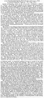
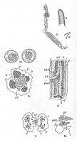

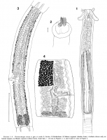

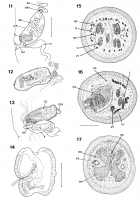
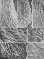
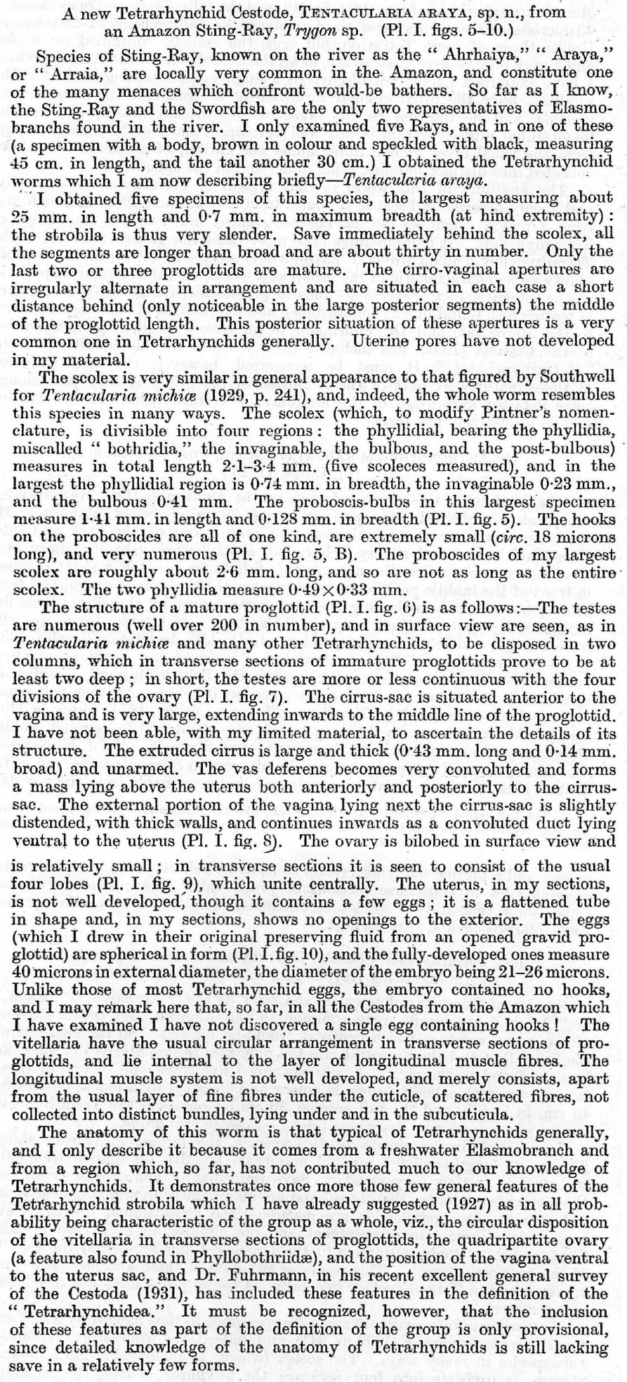
 Fig. 5 (X 12) : the scolex; A ( X 87), portion of a proboscis; B (X 395), spines on tho
proboscides. .
6· ( x 39): dorsal aspect of a gravid proglottid.
7 ( x 56): transverse section of proglottid anterior to o .... ary.
8 ' ( X 87): median ventral portion of · transverse section of proglottid, to show tho
.. position of the vagina ventral to the uterus.
9 (X 87): transverse section through hind end of a young graviu proglottiu in
region of the quadt·ipartite ovary.
10 (X 395): undistorted intra·uterine eggs. Lettering to figures: C.MB. Circular muscle-bands round edges of suckers. OS. Cirrus-sac. DV. Dorsal
excretory canal. LM. Longitudinal muscles. N. Lateral nerve. O. Ovary.
OP. Opening of Bucker. SUBO. Subcuticula. T. Testes. UP. Uterine pore.
U'1.'. Uterus. UTD. Uterine duct. V. Vitellaria. VAG. Vagina. YES. Coils
of vas deferens. VV. Ventral excretory canal.
Fig. 5 (X 12) : the scolex; A ( X 87), portion of a proboscis; B (X 395), spines on tho
proboscides. .
6· ( x 39): dorsal aspect of a gravid proglottid.
7 ( x 56): transverse section of proglottid anterior to o .... ary.
8 ' ( X 87): median ventral portion of · transverse section of proglottid, to show tho
.. position of the vagina ventral to the uterus.
9 (X 87): transverse section through hind end of a young graviu proglottiu in
region of the quadt·ipartite ovary.
10 (X 395): undistorted intra·uterine eggs. Lettering to figures: C.MB. Circular muscle-bands round edges of suckers. OS. Cirrus-sac. DV. Dorsal
excretory canal. LM. Longitudinal muscles. N. Lateral nerve. O. Ovary.
OP. Opening of Bucker. SUBO. Subcuticula. T. Testes. UP. Uterine pore.
U'1.'. Uterus. UTD. Uterine duct. V. Vitellaria. VAG. Vagina. YES. Coils
of vas deferens. VV. Ventral excretory canal.  Campbell et al. (1999)
 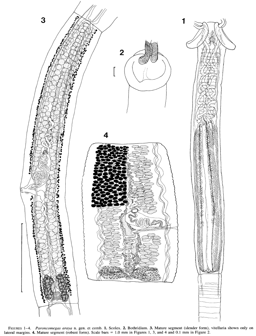 FIGURES 1 -4. Paroncomegas araya n. gen. et comb. 1. Scolex. 2. Bothridium. 3. Mature segment (slender form), vitellaria shown only on lateral margins. 4. Mature segment (robust form). Scale bars = 1.0 mm in Figures 1, 3, and 4 and 0.1 mm in Figure 2.
 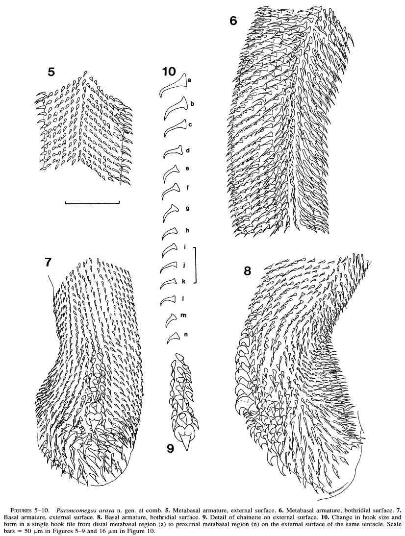 FIGURE5S- 10. Paroncomegas araya n. gen. et comb. 5. Metabasal armature, external surface. 6. Metabasal armature, bothridial surface. 7.
Basal armature, external surface. 8. Basal armature, bothridial surface. 9. Detail of chainette on external surface. 10. Change in hook size and form in a single hook file from distal metabasal region (a) to proximal metabasal region (n) on the external surface of the same tentacle. Scale bars = 50 µm n Figures 5-9 and 16 µm Figure 10.
FIGURE5S- 10. Paroncomegas araya n. gen. et comb. 5. Metabasal armature, external surface. 6. Metabasal armature, bothridial surface. 7.
Basal armature, external surface. 8. Basal armature, bothridial surface. 9. Detail of chainette on external surface. 10. Change in hook size and form in a single hook file from distal metabasal region (a) to proximal metabasal region (n) on the external surface of the same tentacle. Scale bars = 50 µm n Figures 5-9 and 16 µm Figure 10.  FIGURES 11-17. Paroncomegas araya n. gen. et comb. 11. Detail of terminal genitalia. 12. Cirrus sac (frontal section). 13. Detail of terminal genitalia in self-fertilization. 14. Gravid segment. 15. Cross section through mature segment at level of testes. 16. Cross section through mature segment at level of cirrus sac. 17. Cross section through mature segment at level of ovarian isthmus. Scales bars = 0.2 mm in Figures 11-13, 15-17 and 1 mm in Figure 14. Abbreviations: C, cirrus; CS, cirrus sac; DO, dorsal osmoregulatory canal; ESV, external seminal vesicle; GA, genital atrium; MD, male duct; N, nerve cord; 0, ovary; SD, sperm duct; SR, seminal receptacle; T, testis; UT, uterus; V, vagina; VD, vas deferens; VO, ventral osmoregulatory canal; VT, vitelline follicles.
  FIGURES 18-24. SEM of Paroncomegas araya n. gen. et comb. 18. External surface of basal armature showing chainette; note linguiform hooks in central file. 19. Bothridial surface of basal armature, chainette at right; note uncinate form of hooks in files adjacent to central file of large hooks. 20. Distal surface of bothridium; note large spiniform microtriches and small filiform microtriches. 21. Detail of hooks on metabasal armature, bothridial face; note spiniform hooks. 22. Spiniform microtriches on proximal bothridial surface. 23. Metabasal armature, bothridial face; note contrasting shapes of hook tips on internal face (left) and external face (right). 24. Spiniform microtriches on pedunculus scolecis (absent from strobila). Scale bars = 10 [pm in Figures 18, 19, 23 and 1 p.m in Figures 20-22, 24.
 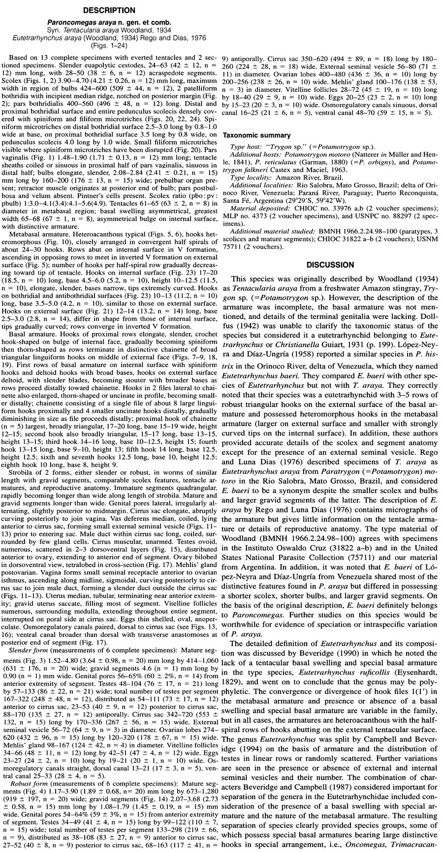 Campbell et al. (1999)
 Best viewed in Firefox


