Cestode Scientific Name
| Species ID | 4641 |
|---|---|
| Order | Trypanorhyncha |
| Family | Eutetrarhynchidae |
| Subfamily | |
| Genus | Oncomegas |
| Species | wageneri |
| Authority | (Linton, 1890) Dollfus, 1929 |
| Taxonomic Status | Valid |
| Valid Name | |
| Synonyms | Rhynchobothrium wageneri Linton, 1890 |
| Genus Record | No |
| Type Species | Yes |
| Verified | Yes |
| Verified By | B. Schaeffner |
| Citation(s) |
Linton, E. 1890. Notes on Entozoa of marine fishes of New England, with descriptions of several new species. Part II. United States Commision of Fish and Fisheries. Part XV. Report of the Commissioner for 1887 15: 718-899. (1011) Download PDFDollfus, R.-P. 1929. Sur les Tetrarhynques. Definition des genres. Bulletin de la Societe Zologique de France 54: 308-342. (4381) Download PDF |
| Redescription |
Toth, L. M., R. A. Campbell, and G. D. Schmidt. 1992. A revision of Oncomegas Dollfus, 1929 (Cestoda: Trypanorhyncha: Eutetrarhynchidae) , the description of two new species and comments on its classification. Systematic Parasitology 22: 167-187. (4264) Download PDF |
| Scientific Name Notes | In some instances, the original authority is cited as Linton (1891), but the appropriate authority is Linton (1890). Original description on page 843 of Linton (1890). Redescribed by Toth et al. (1992), Palm (1995) and Palm (2004). |
Record Data
| Date (MM/DD/YYYY) | Action | User Name |
|---|---|---|
| 11/30/-0001 | Created | I. Beveridge |
| 05/07/2015 | Modified | |
| 10/29/2015 | Modified | N. Arisco |
| 05/13/2016 | Modified | B. Barbeau |
| 06/09/2017 | Modified | K. Herzog |
| 07/27/2017 | Modified | K. Jensen |
| 11/01/2017 | Modified | K. Herzog |
| 12/22/2021 | Modified | B. Barbeau |
Type Host
| Host Class | Elasmobranchii | ||||||
|---|---|---|---|---|---|---|---|
| Host Order | Myliobatiformes | ||||||
| Host Family | Dasyatidae | ||||||
|
Type Host (Literal) |
|
||||||
|
Type Host (Valid) |
|
||||||
| Additional Host(s) | |||||||
| Site in Host | spiral intestine | ||||||
| Host Notes |
Type Locality
| Country | U.S.A. |
|---|---|
| Body of Water | Atlantic Ocean |
| Island(s) | |
| City/Region | Woods Hole, Massachusetts |
| Coordinates | |
| DD Latitude | |
| DD Longitude | |
| Additional Localities | |
| Locality Notes |
Specimens
| Type Material | USNPC 7713 (syntypes) |
|---|---|
| Total Number of Type Specimens | 4 syntypes |
| Voucher Material | |
| Specimen Notes |
Data are given as in original description unless otherwise indicated.
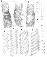
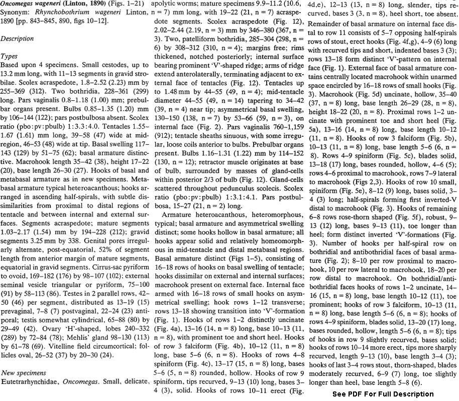
 Figs 1-11. Oncomegas wageneri (Linton, 1890), tentacular armature. 1. Basal armature and proximal metabasal region of tentacle, internal face. 2. Basal armature, antibothridial face. 3. Basal armature and proximal metabasal region of tentacle, external face. 4(a-g). Basal armature hook types, proximal to distal, along one file on internal face. 5(a-f). Basal armature, hook types, proximal to distal, along one file on external face. 6. Proximal metabasal region, internal face. 7. Mid-tentacle region, internal face. 8. Proximal metabasal region, antibothridial face, internal face at left. 9. Antibothridial surface in distal region of tentacle, internal face on left. 10(a-d) Metabasal armature, hook types from one file of internal face, lower metabasal region to tip of tentacle. 11(a-d) Metabasal hooks from one file on external face, lower metabasal region to tip of tentacle.
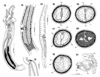
  Figs. 12-15. Oncomegas wageneri (Linton, 1890). 12. Scolex. 13. Mature segment. 14. Gravid segment. 15. Anterior portion of bulb, showing prebulbar organ. Figs 16-21. Oncomegas wageneri (Linton, 1890). 16-20. Transverse sections of mature segment; 16. Anterior to cirrus-sac. 17. Through external seminal vesicle. 18. Through cirrus-sac; note dorsal position of osmoregulatory canals. 19. Posterior to cirrus-sac; note vagina and uterine duct. 20. Through ovarian isthmus, note tetra-loned ovary and osmoregulatory ducts between lobes. 21. Wholemount dorsal view of cirrus sac, external seminal vesicle, and distal vagina. Abbreviations: CS, cirrus-sac; DC, dorsal osmoregulatory canal; ESV, external seminal vesicle; LM, longitudinal muscle bundles; NC, lateral nerve cord; OV, ovary; T, testis; U, uterus; UD, uterine duct; VA, vagina; VC, ventral osmoregulatory canal; VD, vas deferens; VS, vaginal sphincter; VT, vitelline follicle.
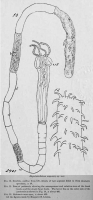
 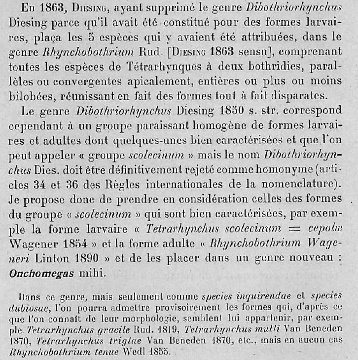 Dollfus (1929) pg. 326
 Best viewed in Firefox


