Line Drawing 1
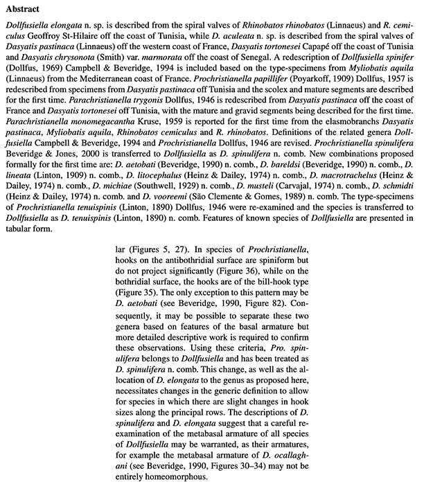
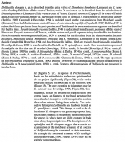
Figures 19. Prochristianella spinulifera n. sp., tentacular armature. 1. Distal region, bothridial surface. 2. Distal region, external surface.
3. Distal region, antibothridial surface. 4. Metabasal... MoreFigures 19. Prochristianella spinulifera n. sp., tentacular armature. 1. Distal region, bothridial surface. 2. Distal region, external surface.
3. Distal region, antibothridial surface. 4. Metabasal region, bothridial surface. 5. Metabasal region, external surface. 6. Metabasal region,
antibothridial surface. 7. Basal region, bothridial surface. 8. Basal region, external surface. 9. Basal region, antibothridial surface. Numerals
indicate hooks of principal rows, beginning on the bothridial surface and extending to the antibothridial surface. Scale-bar: 0.01 mm. |
Line Drawing 2

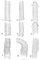
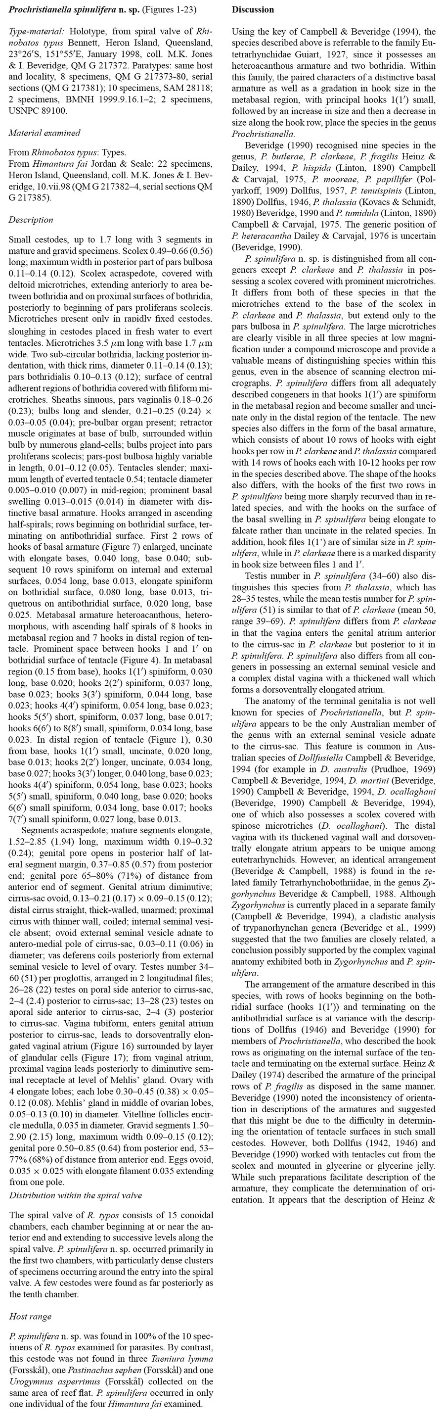
Figures 1017. Prochristianella spinulifera n. sp. 10. Scolex, lateral view. Arrows indicate posterior extent of deltoid microtriches. 11. Scolex,
dorso-ventral view. 12. Entire cestode. 13. Mature s... MoreFigures 1017. Prochristianella spinulifera n. sp. 10. Scolex, lateral view. Arrows indicate posterior extent of deltoid microtriches. 11. Scolex,
dorso-ventral view. 12. Entire cestode. 13. Mature segment. 14. Transverse section of mature segment. 15. Posterior extremity of mature
segment showing terminal genitalia; vitelline follicles are shown on the lateral margins only. 16. Lateral view of genital ducts from genital
atrium, showing dorsoventral elongation of vaginal atrium. 17. Terminal genitalia, showing everted cirrus. Scale-bars: 0.1 mm. Abbreviations:
A, vaginal atrium; C, cirrus-sac; DV, distal vagina; E, external seminal vesicle; PV, posterior vagina; T, testis; U, uterus; V, vitelline glands. |
Photo Micrograph
|
Scanning Electron Micrograph
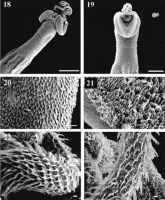
Figures 1823. Prochristianella spinulifera n. sp. Scanning electron micrographs of scolex and hooks. 18. Scolex, lateral view, showing deltoid
microtriches on scolex and external surfaces of bothrid... MoreFigures 1823. Prochristianella spinulifera n. sp. Scanning electron micrographs of scolex and hooks. 18. Scolex, lateral view, showing deltoid
microtriches on scolex and external surfaces of bothridia, density of microtriches on pars pedunculus scolecis diminishing posteriorly. 19.
Scolex, dorsal view, showing filamentous microtriches in V-shaped area on surface of bothridium and deltoid microtriches on pars pedunculus
scolecis. 20. Deltoid microtriches on pars vaginalis scolecis. 21. Surface of bothridium showing filamentous microtriches. 22. Basal swelling of
tentacle, antibothridial surface showing triquetrous hooks. 23. Metabasal region of tentacle, showing ascending rows of spiniform hooks. Scale
bars: 18, 0.1 mm, 19; 0.05 mm, 20; 0.01 mm, 2123; 0.001 mm. |
 Figures 19. Prochristianella spinulifera n. sp., tentacular armature. 1. Distal region, bothridial surface. 2. Distal region, external surface.
3. Distal region, antibothridial surface. 4. Metabasal region, bothridial surface. 5. Metabasal region, external surface. 6. Metabasal region,
antibothridial surface. 7. Basal region, bothridial surface. 8. Basal region, external surface. 9. Basal region, antibothridial surface. Numerals
indicate hooks of principal rows, beginning on the bothridial surface and extending to the antibothridial surface. Scale-bar: 0.01 mm.
Figures 19. Prochristianella spinulifera n. sp., tentacular armature. 1. Distal region, bothridial surface. 2. Distal region, external surface.
3. Distal region, antibothridial surface. 4. Metabasal region, bothridial surface. 5. Metabasal region, external surface. 6. Metabasal region,
antibothridial surface. 7. Basal region, bothridial surface. 8. Basal region, external surface. 9. Basal region, antibothridial surface. Numerals
indicate hooks of principal rows, beginning on the bothridial surface and extending to the antibothridial surface. Scale-bar: 0.01 mm.  Figures 1017. Prochristianella spinulifera n. sp. 10. Scolex, lateral view. Arrows indicate posterior extent of deltoid microtriches. 11. Scolex,
dorso-ventral view. 12. Entire cestode. 13. Mature segment. 14. Transverse section of mature segment. 15. Posterior extremity of mature
segment showing terminal genitalia; vitelline follicles are shown on the lateral margins only. 16. Lateral view of genital ducts from genital
atrium, showing dorsoventral elongation of vaginal atrium. 17. Terminal genitalia, showing everted cirrus. Scale-bars: 0.1 mm. Abbreviations:
A, vaginal atrium; C, cirrus-sac; DV, distal vagina; E, external seminal vesicle; PV, posterior vagina; T, testis; U, uterus; V, vitelline glands.
Figures 1017. Prochristianella spinulifera n. sp. 10. Scolex, lateral view. Arrows indicate posterior extent of deltoid microtriches. 11. Scolex,
dorso-ventral view. 12. Entire cestode. 13. Mature segment. 14. Transverse section of mature segment. 15. Posterior extremity of mature
segment showing terminal genitalia; vitelline follicles are shown on the lateral margins only. 16. Lateral view of genital ducts from genital
atrium, showing dorsoventral elongation of vaginal atrium. 17. Terminal genitalia, showing everted cirrus. Scale-bars: 0.1 mm. Abbreviations:
A, vaginal atrium; C, cirrus-sac; DV, distal vagina; E, external seminal vesicle; PV, posterior vagina; T, testis; U, uterus; V, vitelline glands.  Figures 1823. Prochristianella spinulifera n. sp. Scanning electron micrographs of scolex and hooks. 18. Scolex, lateral view, showing deltoid
microtriches on scolex and external surfaces of bothridia, density of microtriches on pars pedunculus scolecis diminishing posteriorly. 19.
Scolex, dorsal view, showing filamentous microtriches in V-shaped area on surface of bothridium and deltoid microtriches on pars pedunculus
scolecis. 20. Deltoid microtriches on pars vaginalis scolecis. 21. Surface of bothridium showing filamentous microtriches. 22. Basal swelling of
tentacle, antibothridial surface showing triquetrous hooks. 23. Metabasal region of tentacle, showing ascending rows of spiniform hooks. Scale
bars: 18, 0.1 mm, 19; 0.05 mm, 20; 0.01 mm, 2123; 0.001 mm.
Figures 1823. Prochristianella spinulifera n. sp. Scanning electron micrographs of scolex and hooks. 18. Scolex, lateral view, showing deltoid
microtriches on scolex and external surfaces of bothridia, density of microtriches on pars pedunculus scolecis diminishing posteriorly. 19.
Scolex, dorsal view, showing filamentous microtriches in V-shaped area on surface of bothridium and deltoid microtriches on pars pedunculus
scolecis. 20. Deltoid microtriches on pars vaginalis scolecis. 21. Surface of bothridium showing filamentous microtriches. 22. Basal swelling of
tentacle, antibothridial surface showing triquetrous hooks. 23. Metabasal region of tentacle, showing ascending rows of spiniform hooks. Scale
bars: 18, 0.1 mm, 19; 0.05 mm, 20; 0.01 mm, 2123; 0.001 mm.