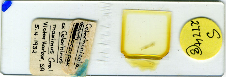
Line Drawing 1
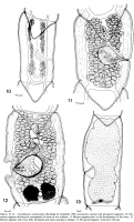
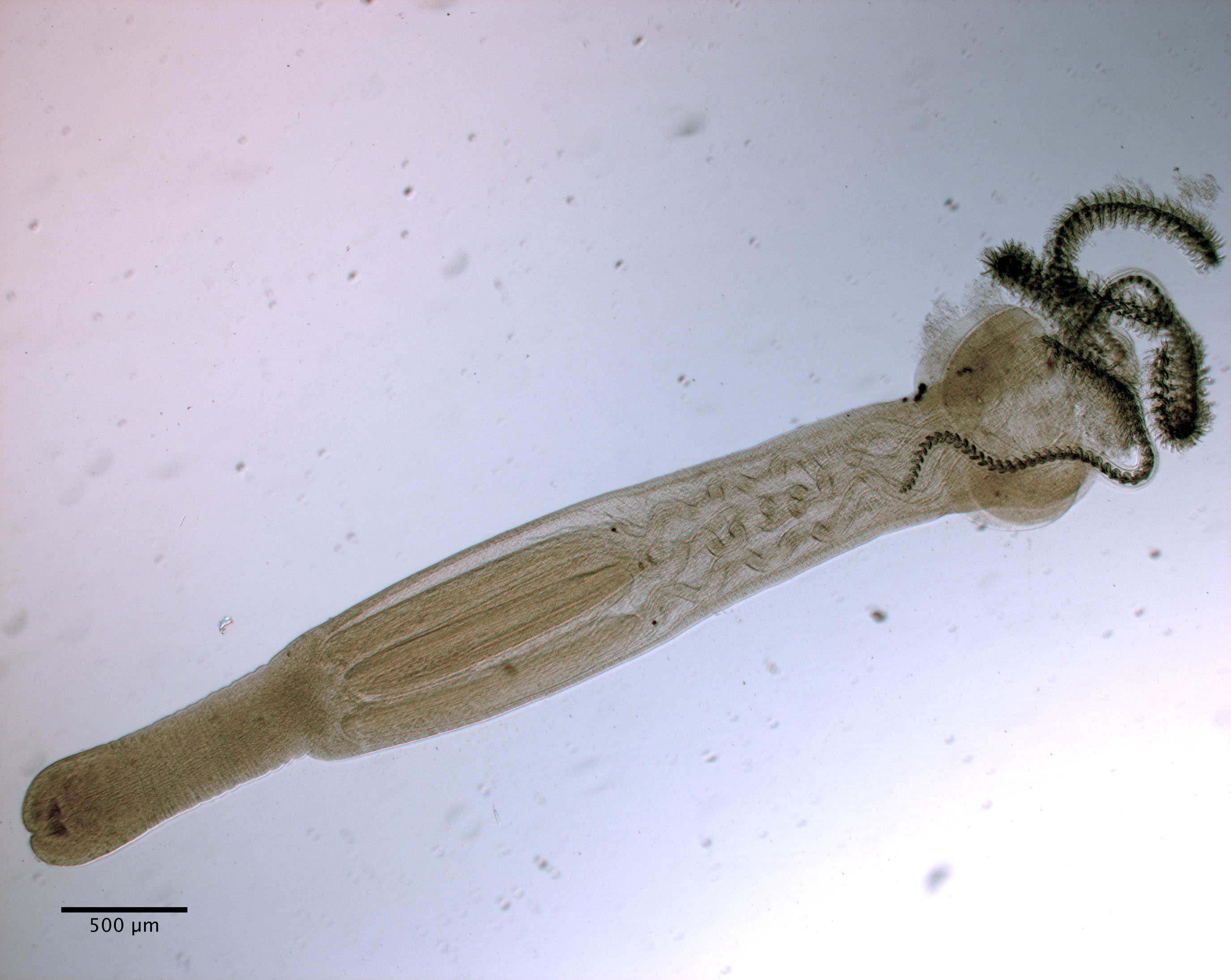
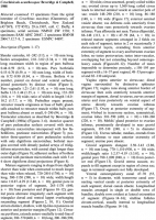
Figures 1013. Cetorhinicola acanthocapax Beveridge & Campbell, 1988, pre-mature, mature and pre-gravid segments. 10. Premature
segment showing the arrangement of testes in two columns. 11. Mature se... MoreFigures 1013. Cetorhinicola acanthocapax Beveridge & Campbell, 1988, pre-mature, mature and pre-gravid segments. 10. Premature
segment showing the arrangement of testes in two columns. 11. Mature segment prior to the development of the ovary. 12.
Mature segment with ovary fully developed and uterus starting to enlarge. 13. Pre-gravid segment. Scale-bars: 100 lm. |
Line Drawing 2
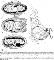
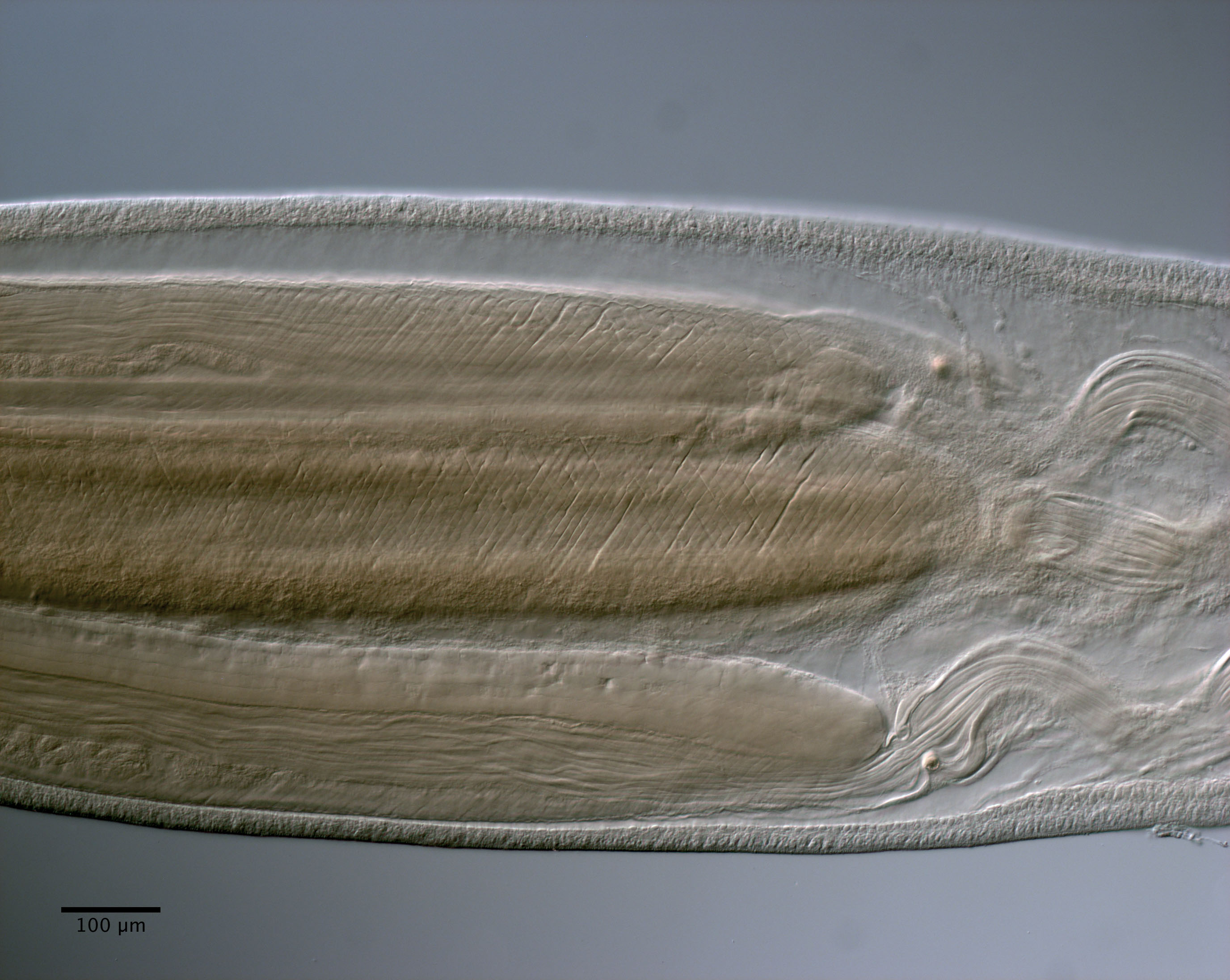
Figures 1417. Cetorhinicola acanthocapax Beveridge & Campbell, 1988, transverse sections through mature segments and dorsoventral
illustration of the terminal genital ducts. 14. Transverse section t... MoreFigures 1417. Cetorhinicola acanthocapax Beveridge & Campbell, 1988, transverse sections through mature segments and dorsoventral
illustration of the terminal genital ducts. 14. Transverse section through the anterior region of a segment, including the lappets
of the preceding segment. 15. Transverse section of a mature segment through the genital atrium showing the cirrus-sac. 16.
Transverse section of a mature segment at the level of the ovary and Mehlis gland. 17. Dorso-ventral view of the genital ducts.
Abbreviations: c, cirrus; cs, cirrus-sac; ga, genital atrium; is, internal seminal vesicle; l, lappet; lm, longitudinal muscle; mg, Mehlis
gland; o, ovary; sp, sphincter; t, testis; u, uterus; v, vagina; vd, vas deferens; vi, vitelline follicle; vo, ventral osmoregulatory canal.
Scale-bars: 100 lm |
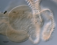
Photo Micrograph
|
Scanning Electron Micrograph
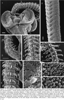
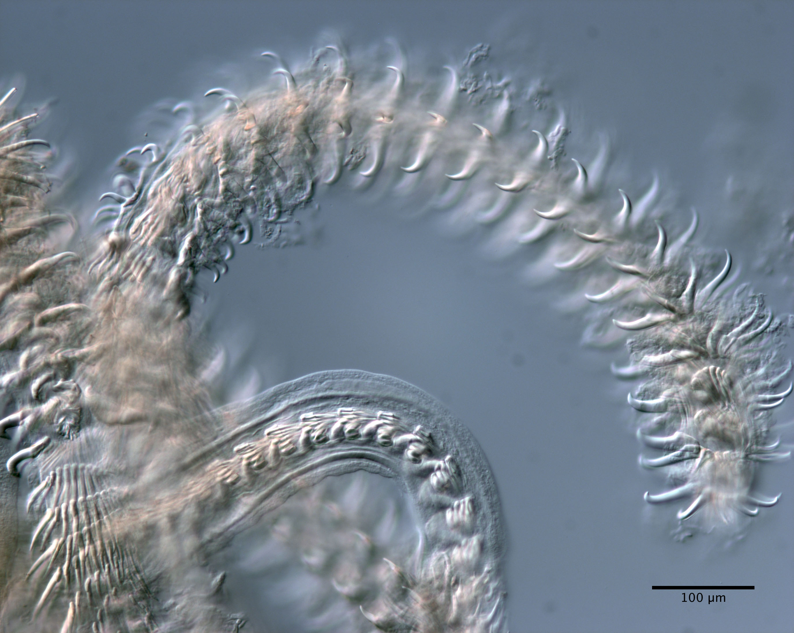
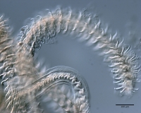
Figures 19. Cetorhinicola acanthocapax Beveridge & Campbell, 1988, scanning electron micrographs of the scolex, strobila, tentacles
and microtriches. 1. Scolex showing bothria joined at their poster... MoreFigures 19. Cetorhinicola acanthocapax Beveridge & Campbell, 1988, scanning electron micrographs of the scolex, strobila, tentacles
and microtriches. 1. Scolex showing bothria joined at their posterior margin by a distinct velum. 2. Immature segments showing
lappets overhanging succeeding segments. 3. Basal armature of tentacle, external surface on left hand side. 4. Metabasal
armature, external surface showing space between hook files 8 and 8. 5. Metabasal armature, oblique view of the internal surface.
6. Metabasal armature, antibothrial surface. 7. Pectinate microtriches scattered among short digitiform microtriches on the anterior
part of the pedunculus scolecis immediately posterior to the bothria. 8. Pectinate microtriches on the adherent surface of the
bothrium. 9. Trigitate microtriches on the outer margin of the bothrium. Abbreviations v, velum. Scale-bars: 13,79, 100 lm; 4
6,1 lm. |
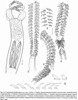


 Figs 1-6. Cetorhinicola acanthocapax n. g., n. sp. 1. Scolex. 2. Tentacle, basal and metabasal regions, internal surface; bothridial surface
to right hand side. 3. Tentacle, basal and metabasal regions, antibothridial surface. 4. Tentacle, basal and metabasal regions, external
surface, bothridial surface to right hand side. 5. Tentacle, metabasal region, external surface; bothridial surface to right hand side. 6.
Hooks 1-7 of metabasal region in profile; enlarged hooks of antibothridial (a) and bothridial (b) surfaces of tentacle in profile. Scale
lines 0.1mm.
Figs 1-6. Cetorhinicola acanthocapax n. g., n. sp. 1. Scolex. 2. Tentacle, basal and metabasal regions, internal surface; bothridial surface
to right hand side. 3. Tentacle, basal and metabasal regions, antibothridial surface. 4. Tentacle, basal and metabasal regions, external
surface, bothridial surface to right hand side. 5. Tentacle, metabasal region, external surface; bothridial surface to right hand side. 6.
Hooks 1-7 of metabasal region in profile; enlarged hooks of antibothridial (a) and bothridial (b) surfaces of tentacle in profile. Scale
lines 0.1mm. 
 Figures 1013. Cetorhinicola acanthocapax Beveridge & Campbell, 1988, pre-mature, mature and pre-gravid segments. 10. Premature
segment showing the arrangement of testes in two columns. 11. Mature segment prior to the development of the ovary. 12.
Mature segment with ovary fully developed and uterus starting to enlarge. 13. Pre-gravid segment. Scale-bars: 100 lm.
Figures 1013. Cetorhinicola acanthocapax Beveridge & Campbell, 1988, pre-mature, mature and pre-gravid segments. 10. Premature
segment showing the arrangement of testes in two columns. 11. Mature segment prior to the development of the ovary. 12.
Mature segment with ovary fully developed and uterus starting to enlarge. 13. Pre-gravid segment. Scale-bars: 100 lm.  Figures 1417. Cetorhinicola acanthocapax Beveridge & Campbell, 1988, transverse sections through mature segments and dorsoventral
illustration of the terminal genital ducts. 14. Transverse section through the anterior region of a segment, including the lappets
of the preceding segment. 15. Transverse section of a mature segment through the genital atrium showing the cirrus-sac. 16.
Transverse section of a mature segment at the level of the ovary and Mehlis gland. 17. Dorso-ventral view of the genital ducts.
Abbreviations: c, cirrus; cs, cirrus-sac; ga, genital atrium; is, internal seminal vesicle; l, lappet; lm, longitudinal muscle; mg, Mehlis
gland; o, ovary; sp, sphincter; t, testis; u, uterus; v, vagina; vd, vas deferens; vi, vitelline follicle; vo, ventral osmoregulatory canal.
Scale-bars: 100 lm
Figures 1417. Cetorhinicola acanthocapax Beveridge & Campbell, 1988, transverse sections through mature segments and dorsoventral
illustration of the terminal genital ducts. 14. Transverse section through the anterior region of a segment, including the lappets
of the preceding segment. 15. Transverse section of a mature segment through the genital atrium showing the cirrus-sac. 16.
Transverse section of a mature segment at the level of the ovary and Mehlis gland. 17. Dorso-ventral view of the genital ducts.
Abbreviations: c, cirrus; cs, cirrus-sac; ga, genital atrium; is, internal seminal vesicle; l, lappet; lm, longitudinal muscle; mg, Mehlis
gland; o, ovary; sp, sphincter; t, testis; u, uterus; v, vagina; vd, vas deferens; vi, vitelline follicle; vo, ventral osmoregulatory canal.
Scale-bars: 100 lm 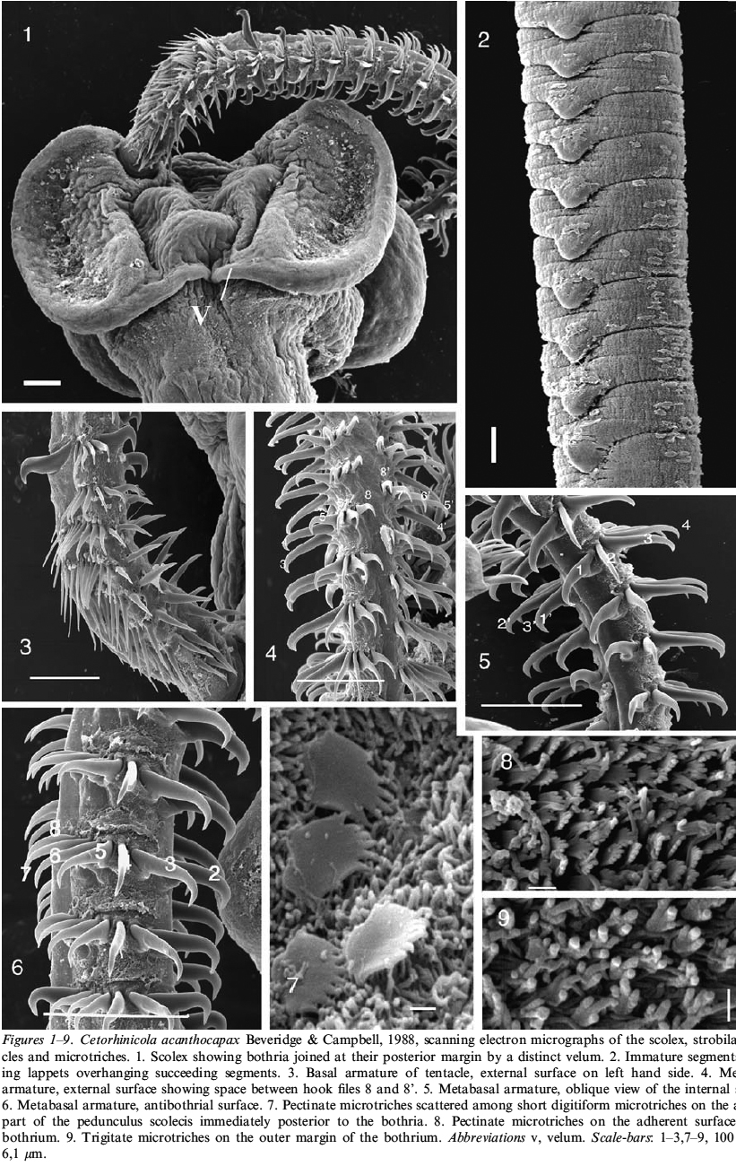 Figures 19. Cetorhinicola acanthocapax Beveridge & Campbell, 1988, scanning electron micrographs of the scolex, strobila, tentacles
and microtriches. 1. Scolex showing bothria joined at their posterior margin by a distinct velum. 2. Immature segments showing
lappets overhanging succeeding segments. 3. Basal armature of tentacle, external surface on left hand side. 4. Metabasal
armature, external surface showing space between hook files 8 and 8. 5. Metabasal armature, oblique view of the internal surface.
6. Metabasal armature, antibothrial surface. 7. Pectinate microtriches scattered among short digitiform microtriches on the anterior
part of the pedunculus scolecis immediately posterior to the bothria. 8. Pectinate microtriches on the adherent surface of the
bothrium. 9. Trigitate microtriches on the outer margin of the bothrium. Abbreviations v, velum. Scale-bars: 13,79, 100 lm; 4
6,1 lm.
Figures 19. Cetorhinicola acanthocapax Beveridge & Campbell, 1988, scanning electron micrographs of the scolex, strobila, tentacles
and microtriches. 1. Scolex showing bothria joined at their posterior margin by a distinct velum. 2. Immature segments showing
lappets overhanging succeeding segments. 3. Basal armature of tentacle, external surface on left hand side. 4. Metabasal
armature, external surface showing space between hook files 8 and 8. 5. Metabasal armature, oblique view of the internal surface.
6. Metabasal armature, antibothrial surface. 7. Pectinate microtriches scattered among short digitiform microtriches on the anterior
part of the pedunculus scolecis immediately posterior to the bothria. 8. Pectinate microtriches on the adherent surface of the
bothrium. 9. Trigitate microtriches on the outer margin of the bothrium. Abbreviations v, velum. Scale-bars: 13,79, 100 lm; 4
6,1 lm.