Cestode Scientific Name
| Species ID | 459 |
|---|---|
| Order | Cyclophyllidea |
| Family | Davaineidae |
| Subfamily | |
| Genus | Cotugnia |
| Species | margareta |
| Authority | Beddard, 1916 |
| Taxonomic Status | Valid |
| Valid Name | |
| Synonyms | |
| Genus Record | No |
| Type Species | No |
| Verified | No |
| Verified By | |
| Citation(s) |
Beddard, F. E. 1916. On two new species of cestodes belonging respectively to the genera Linstowia and Cotugnia. Proceedings of the Zoological Society of London 1916(4): 695-706. (2806) Download PDF |
| Redescription | |
| Scientific Name Notes |
Record Data
| Date (MM/DD/YYYY) | Action | User Name |
|---|---|---|
| 01/09/2006 | Created | Healy, Georgiev |
| 10/15/2013 | Modified | |
| 05/10/2016 | Modified | B. Barbeau |
Type Host
| Host Class | |||||||
|---|---|---|---|---|---|---|---|
| Host Order | |||||||
| Host Family | |||||||
|
Type Host (Literal) |
|
||||||
|
Type Host (Valid) |
|
||||||
| Additional Host(s) | |||||||
| Site in Host | gut | ||||||
| Host Notes |
Type Locality
| Country | [not given] |
|---|---|
| Body of Water | |
| Island(s) | |
| City/Region | |
| Coordinates | |
| DD Latitude | |
| DD Longitude | |
| Additional Localities | |
| Locality Notes | Locality not given but likely obtained from the Zoological Gardens in London, England |
Specimens
| Type Material | [not given] |
|---|---|
| Total Number of Type Specimens | |
| Voucher Material | |
| Specimen Notes |
Data are given as in original description unless otherwise indicated.
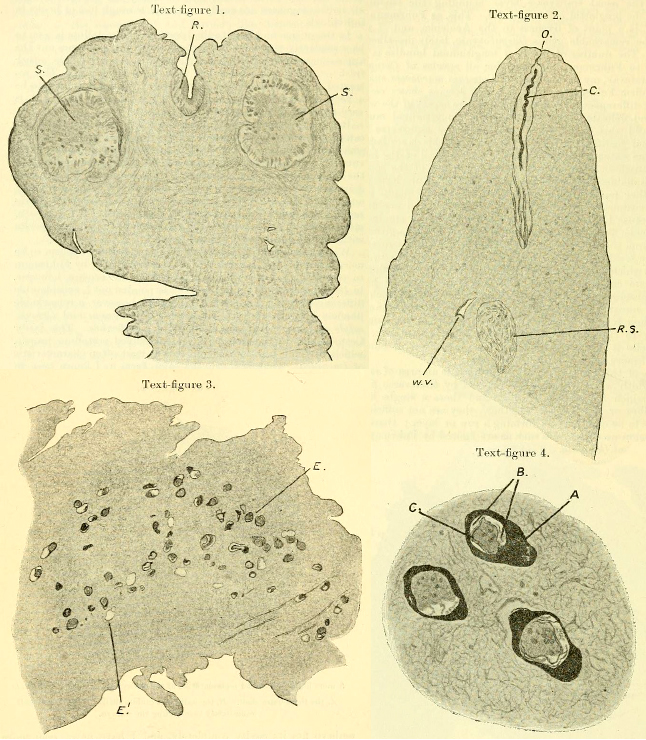 Text-figure 1. Longitudinal section through scolex of Cotugnia margareta. R. rostellum, in which the hooks are not shown. S. suckers. Text-figure 2. Cirrus-sac of Cotugnia margareta. C. cirrus lying within the elongated cirrus-sac. R.S. receptaculum seminis. W.V. ventral water-vascular tube. O. orifice of cirrus-sac. Text-figure 3. Section (horizontal) through two adjacent segments of Cotugnia margareta, showing the embyros scattered through the medullary parenchyma. E. embryos. E1. spaces from which an embryo has fallen out. Text-figure 4. A more highly magnified represenation of the embryos of Cotugnia margareta. A. the thick outer shell. B. the indistinct middle shelf. C. the inner shell immediately surrounding the embryo.
 Best viewed in Firefox


