Cestode Scientific Name
| Species ID | 37 |
|---|---|
| Order | Cathetocephalidea |
| Family | Cathetocephalidae |
| Subfamily | |
| Genus | Cathetocephalus |
| Species | thatcheri |
| Authority | Dailey & Overstreet, 1973 |
| Taxonomic Status | Valid |
| Valid Name | |
| Synonyms | |
| Genus Record | No |
| Type Species | Yes |
| Verified | No |
| Verified By | |
| Citation(s) |
Dailey, M. D. and R. M. Overstreet. 1973. Cathetocephalus thatcheri gen. et sp. n. (Tetraphyllidea: Cathetocephalidae fam. n.) from the bull shark: A species demonstrating multistrobilization. Journal of Parasitology 59: 469-473. (154) Download PDF |
| Redescription | |
| Scientific Name Notes |
Record Data
| Date (MM/DD/YYYY) | Action | User Name |
|---|---|---|
| 09/09/2006 | Created | J. N. Caira |
| 02/04/2010 | Modified | |
| 01/07/2016 | Modified | |
| 05/10/2016 | Modified | B. Barbeau |
Type Host
| Host Class | |||||||
|---|---|---|---|---|---|---|---|
| Host Order | Carcharhiniformes | ||||||
| Host Family | Carcharhinidae | ||||||
|
Type Host (Literal) |
|
||||||
|
Type Host (Valid) |
|
||||||
| Additional Host(s) | |||||||
| Site in Host | spiral valve | ||||||
| Host Notes |
Type Locality
| Country | U.S.A. |
|---|---|
| Body of Water | Galveston Bay |
| Island(s) | |
| City/Region | Texas |
| Coordinates | |
| DD Latitude | |
| DD Longitude | |
| Additional Localities | additional locality: off of Miami, Florida, U. S. A. |
| Locality Notes |
Specimens
| Type Material | USNPC No. 72302 (holotype) USNPC No. 72302 |
|---|---|
| Total Number of Type Specimens | 15 |
| Voucher Material | Based on 15 specimens |
| Specimen Notes |
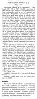
Data are given as in original description unless otherwise indicated.


 FIGURES 1-3. Cathetocephalus thatcheri n. sp. 1. Transverse scolex, ventral view. 2. Mature proglottid. 3. Diagrammatic illustration of multistrobilate form, dorsal view (approximately x7). C, cirrus; M, Mehlis gland; O, ovary; U, uterus; T, testis; V, vitelline follicle; VA, vagina; VD, vas deferens.
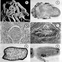
 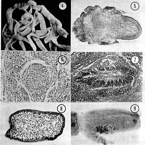 FIGURES 4-9. 4. Multistrobilate form, ventral view (x7). 5. Swirling pattern of strobila formation site in scolex of multistrobilate form (x77). 6. Isolation of strobila formation site in scolex of multistrobilate form (x214). 7. Isolation site of strobila showing cellular differentiation in scolex of multistrobilate form (x125). 8. Anterior region of strobila showing welldefined cortical and medullary regions (x255). 9. Budding process seen in mature segment of unistrobilate form (x22).
 Best viewed in Firefox


