Line Drawing 1
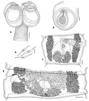
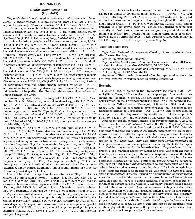
FIGURES 48. G. argentinense n. gen. et n. sp. (4) Scolex, bar = 400 µm. (5) Detail of bothridium in apical view, bar = 200 µm. (6) Eggs, bar = 25 µm. (7) Gravid segment, bar = 200 µm. (8) Mature segm... MoreFIGURES 48. G. argentinense n. gen. et n. sp. (4) Scolex, bar = 400 µm. (5) Detail of bothridium in apical view, bar = 200 µm. (6) Eggs, bar = 25 µm. (7) Gravid segment, bar = 200 µm. (8) Mature segment, bar = 200 µm. |
Line Drawing 2

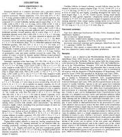
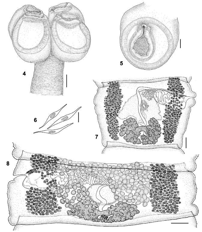
FIGURES 914. G. argentinense n. gen. et n. sp. (9) Entire worm, bar = 400 µm. (1014) Cross sections of gravid segment, bar = 200 µm. (10) At level of testes anterior to cirrus sac. (11) At level of ... MoreFIGURES 914. G. argentinense n. gen. et n. sp. (9) Entire worm, bar = 400 µm. (1014) Cross sections of gravid segment, bar = 200 µm. (10) At level of testes anterior to cirrus sac. (11) At level of genital pore. (12) At level of uterine pore. (13) At level of ovarian isthmus. (14) At level of Mehlis gland. Abbreviations: cs, cirrus sac; dod, dorsal osmoregulatory duct; mg, Mehlis gland; ov, ovary; t, testis; u, uterus; up, uterine pore; vd, vas deferens; vf, vitelline follicle; vs, vaginal sphincter; vod, ventral osmoregulatory duct. |
Photo Micrograph
|
Scanning Electron Micrograph

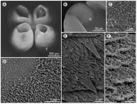
FIGURES 1519. G. argentinense n. gen. et n. sp., scanning electron micrographs. (15) Scolex, lateral view, bar = 500 µm. (16) Scolex, apical view, bar = 500 µm. (17) Detail of accessory sucker, bar =... MoreFIGURES 1519. G. argentinense n. gen. et n. sp., scanning electron micrographs. (15) Scolex, lateral view, bar = 500 µm. (16) Scolex, apical view, bar = 500 µm. (17) Detail of accessory sucker, bar = 100 µm. (18) Detail of scutes on the surface of germinative zone, bar = 50 µm. (19) Detail of microtriches on scutes, bar = 2µm. |
 FIGURES 48. G. argentinense n. gen. et n. sp. (4) Scolex, bar = 400 µm. (5) Detail of bothridium in apical view, bar = 200 µm. (6) Eggs, bar = 25 µm. (7) Gravid segment, bar = 200 µm. (8) Mature segment, bar = 200 µm.
 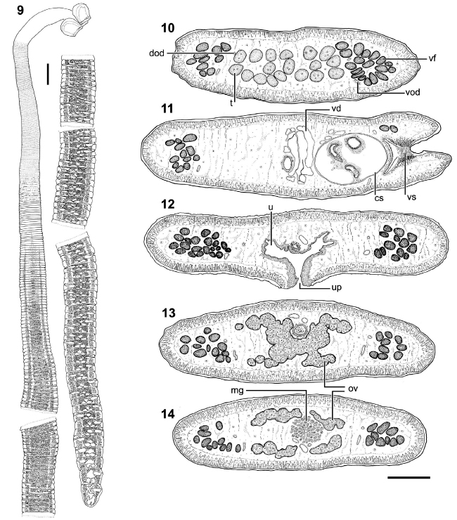 FIGURES 914. G. argentinense n. gen. et n. sp. (9) Entire worm, bar = 400 µm. (1014) Cross sections of gravid segment, bar = 200 µm. (10) At level of testes anterior to cirrus sac. (11) At level of genital pore. (12) At level of uterine pore. (13) At level of ovarian isthmus. (14) At level of Mehlis gland. Abbreviations: cs, cirrus sac; dod, dorsal osmoregulatory duct; mg, Mehlis gland; ov, ovary; t, testis; u, uterus; up, uterine pore; vd, vas deferens; vf, vitelline follicle; vs, vaginal sphincter; vod, ventral osmoregulatory duct.
 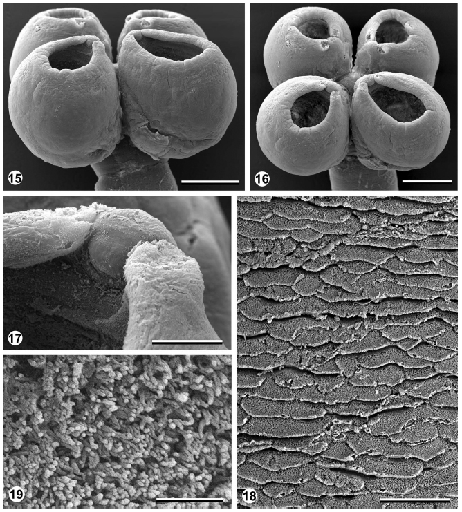 FIGURES 1519. G. argentinense n. gen. et n. sp., scanning electron micrographs. (15) Scolex, lateral view, bar = 500 µm. (16) Scolex, apical view, bar = 500 µm. (17) Detail of accessory sucker, bar = 100 µm. (18) Detail of scutes on the surface of germinative zone, bar = 50 µm. (19) Detail of microtriches on scutes, bar = 2µm.
 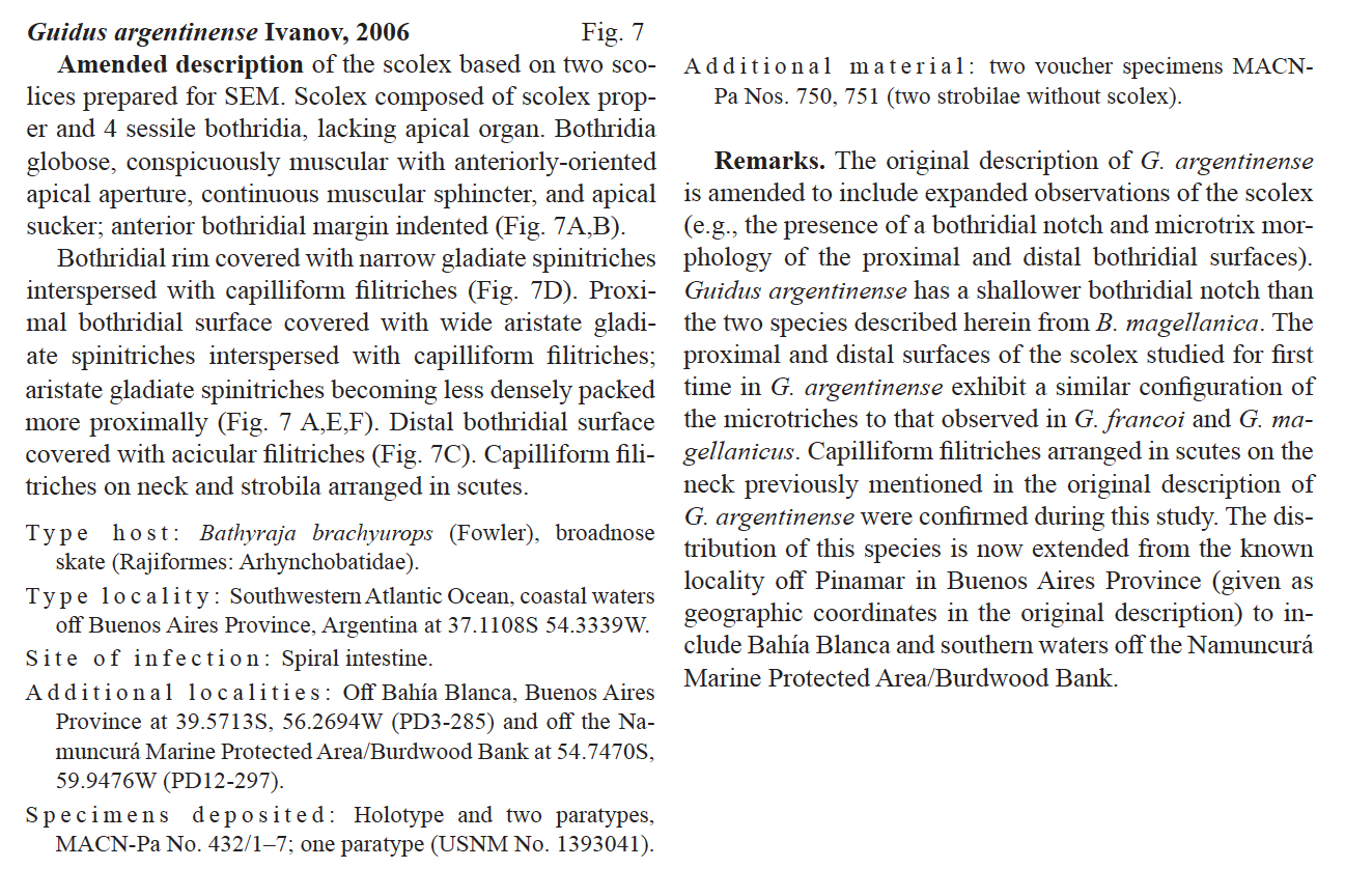 From Menoret & Ivanov, 2021 (Cit# 7321)
 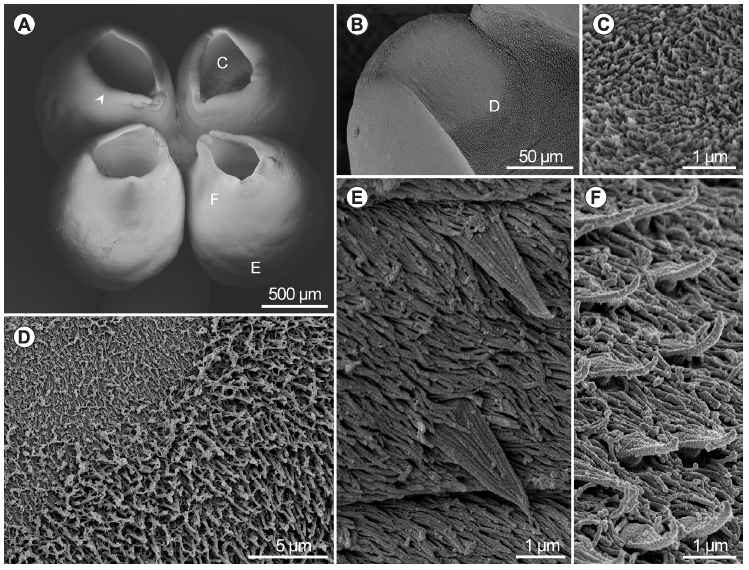 From Menoret & Ivanov, 2021 (Cit# 7321); Fig. 7. Guidus argentinense Ivanov, 2006 from Bathyraja brachyurops (Fowler), scanning electron micrographs. A scolex, small letters indicate locations of detail shown in C, E and F, arrow indicates marginal indentation; B apical sucker, small letter indicates location of detail shown in D; C distal bothridial surface, acicular filitriches; D detail of apical sucker and surrounding area; E proximal bothridial surface near proximal bothridial extreme, wide aristate gladiate spinitriches and capilliform filitriches; F proximal bothridial surface near apical rim showing wide aristate gladiate spinitriches with pad-shaped bases

