Line Drawing 1
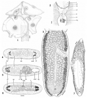
From Ivanov, 2008 (Cit #4439). FIGURES 17. Orygmatobothrium schmittii Suriano and Labriola, 2001. (1) Scolex, scale bar=1 mm. (2) Detail of ootype region, scale bar=100 µm. (3) Last mature proglottid... MoreFrom Ivanov, 2008 (Cit #4439). FIGURES 17. Orygmatobothrium schmittii Suriano and Labriola, 2001. (1) Scolex, scale bar=1 mm. (2) Detail of ootype region, scale bar=100 µm. (3) Last mature proglottid, scale bar=200 µm. (46) Cross sections of mature proglottid, scale bar=100 µm. (4) At level of testes anterior to cirrus sac. (5) At level of genital pore. (6) At level of ovarian isthmus. (7) Detached gravid proglottid, scale bar=600 µm.
Abbreviations: cs, cirrus sac; lm, longitudinal musculature; mg, Mehliss gland; nc, nerve cord; ov, ovary; sr, seminal receptacle; t, testis; u,
uterus; ud, uterine duct; vg, vagina; vd, vas deferens; vf, vitelline follicle; vod, ventral osmoregulatory duct. |
Line Drawing 2
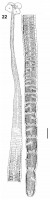
From Ivanov, 2008 (Cit #4439). FIGURE 22. Entire worms, scale bar=1 mm. Orygmatobothrium schmittii Suriano and Labriola, 2001. |
Photo Micrograph
|
Scanning Electron Micrograph
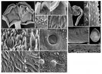
From Ivanov, 2008 (Cit #4439). FIGURES 816. Orygmatobothrium schmittii Suriano and Labriola, 2001, scanning electron micrographs. (8) Scolex, scale bar=100 µm. (9) Accessory sucker at anterior margin... MoreFrom Ivanov, 2008 (Cit #4439). FIGURES 816. Orygmatobothrium schmittii Suriano and Labriola, 2001, scanning electron micrographs. (8) Scolex, scale bar=100 µm. (9) Accessory sucker at anterior margin of bothridium, arrow indicates cleft with overlapping margins, scale bar=10 µm. (10) Detail of cephalic peduncle and germinative zone, scale bar=25 µm. (11) Detail of microtriches on cephalic peduncle, scale bar=2.5 µm. (12) Detail of microtriches on scolex proper, scale bar=1 µm. (13) Distal bothridial surface, scale bar=2 µm; inset shows enlarged view of microtriches,
scale bar=1 µm. (14) Glandulomuscular organ on distal bothridial surface; arrows indicate the presence of rounded projections with cilia
magnified in Figure 16, scale bar=20 µm. (15) Proximal bothridial surface, scale bar=2 µm; inset shows enlarged view of microtriches, arrows indicate position of projections in trifid microtriches, scale bar=1 µm. (16) Detail of a rounded projection with central cilium and filiform microtriches on outer surface of glandulomuscular organ, scale bar=2 µm. (17) Everted cirrus, scale bar=100 µm; inset (superior corner) shows enlarged view of microtriches covering cirrus basal swelling, scale bar=2 µm; inset (inferior corner) shows enlarged view of microtriches covering cirrus distal slender part, scale bar=2 µm. (18) Surface of mature proglottid showing papillae
surrounding genital pore, scale bar=10 µm. (19) Detail of microtriches forming scutes on germinative zone, scale bar=2 µm. (20) Egg, scale bar=5 µm. (21) Detail of microtriches on surface of mature proglottids, scale bar=2 µm. |

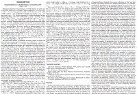
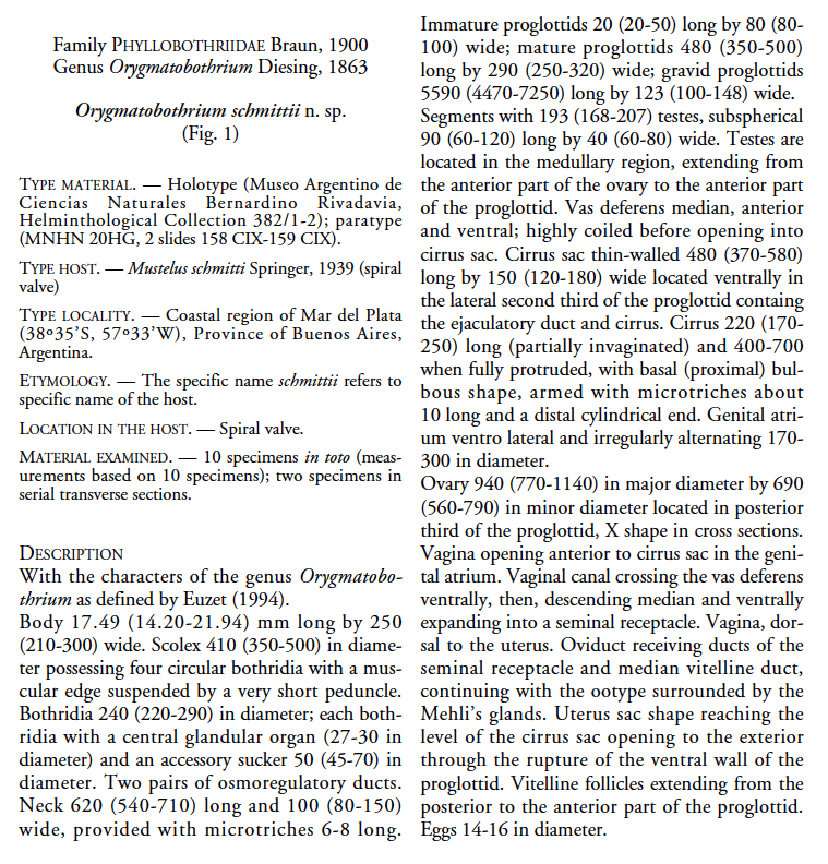
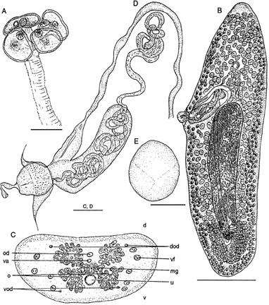 FIG. 1. Orygmatobothrium schmittii n. sp.; A, scolex; B, gravid proglottid (ventral view); C, cross section throught proglottid at level
of ovary; D, detail of terminal genitalia with everted cirrus (lateral view); E, egg. Abbreviations: d, dorsal; v, ventral; o, ovary;
od, oviduct; dod, dorsal osmorregulatory duct; vod, ventral osmoregulatory duct; mg, mehlis glands; va, vagin; vf, vitelline follicles;
u, uterus. Scale bars: A-C, 0.2 mm; B, 1 mm; D, 0.05 mm; E, 0.01 mm.
FIG. 1. Orygmatobothrium schmittii n. sp.; A, scolex; B, gravid proglottid (ventral view); C, cross section throught proglottid at level
of ovary; D, detail of terminal genitalia with everted cirrus (lateral view); E, egg. Abbreviations: d, dorsal; v, ventral; o, ovary;
od, oviduct; dod, dorsal osmorregulatory duct; vod, ventral osmoregulatory duct; mg, mehlis glands; va, vagin; vf, vitelline follicles;
u, uterus. Scale bars: A-C, 0.2 mm; B, 1 mm; D, 0.05 mm; E, 0.01 mm. 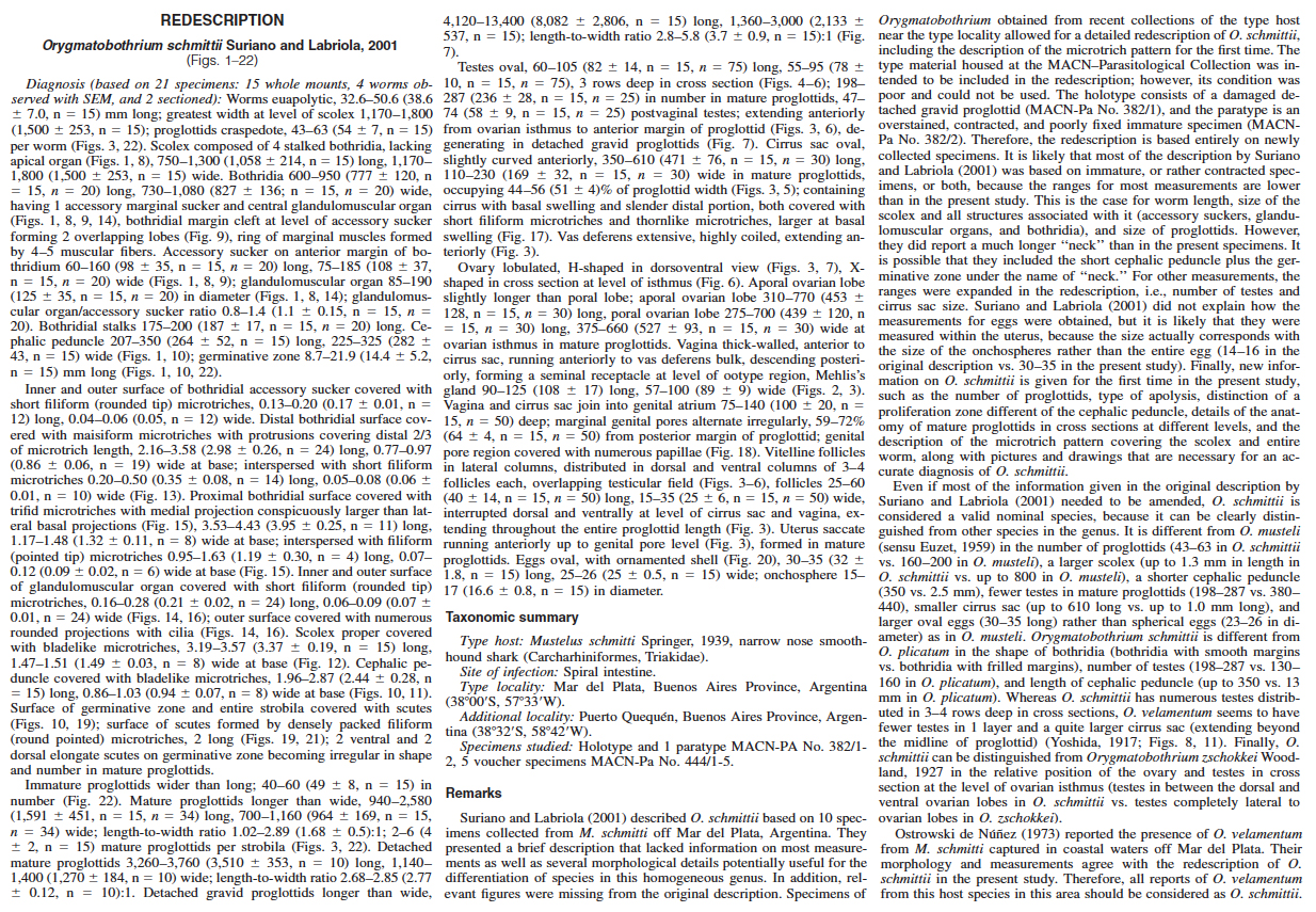 From Ivanov, 2008 (Cit #4439)
  From Ivanov, 2008 (Cit #4439). FIGURES 17. Orygmatobothrium schmittii Suriano and Labriola, 2001. (1) Scolex, scale bar=1 mm. (2) Detail of ootype region, scale bar=100 µm. (3) Last mature proglottid, scale bar=200 µm. (46) Cross sections of mature proglottid, scale bar=100 µm. (4) At level of testes anterior to cirrus sac. (5) At level of genital pore. (6) At level of ovarian isthmus. (7) Detached gravid proglottid, scale bar=600 µm.
Abbreviations: cs, cirrus sac; lm, longitudinal musculature; mg, Mehliss gland; nc, nerve cord; ov, ovary; sr, seminal receptacle; t, testis; u,
uterus; ud, uterine duct; vg, vagina; vd, vas deferens; vf, vitelline follicle; vod, ventral osmoregulatory duct.
From Ivanov, 2008 (Cit #4439). FIGURES 17. Orygmatobothrium schmittii Suriano and Labriola, 2001. (1) Scolex, scale bar=1 mm. (2) Detail of ootype region, scale bar=100 µm. (3) Last mature proglottid, scale bar=200 µm. (46) Cross sections of mature proglottid, scale bar=100 µm. (4) At level of testes anterior to cirrus sac. (5) At level of genital pore. (6) At level of ovarian isthmus. (7) Detached gravid proglottid, scale bar=600 µm.
Abbreviations: cs, cirrus sac; lm, longitudinal musculature; mg, Mehliss gland; nc, nerve cord; ov, ovary; sr, seminal receptacle; t, testis; u,
uterus; ud, uterine duct; vg, vagina; vd, vas deferens; vf, vitelline follicle; vod, ventral osmoregulatory duct. 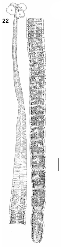 From Ivanov, 2008 (Cit #4439). FIGURE 22. Entire worms, scale bar=1 mm. Orygmatobothrium schmittii Suriano and Labriola, 2001.
  From Ivanov, 2008 (Cit #4439). FIGURES 816. Orygmatobothrium schmittii Suriano and Labriola, 2001, scanning electron micrographs. (8) Scolex, scale bar=100 µm. (9) Accessory sucker at anterior margin of bothridium, arrow indicates cleft with overlapping margins, scale bar=10 µm. (10) Detail of cephalic peduncle and germinative zone, scale bar=25 µm. (11) Detail of microtriches on cephalic peduncle, scale bar=2.5 µm. (12) Detail of microtriches on scolex proper, scale bar=1 µm. (13) Distal bothridial surface, scale bar=2 µm; inset shows enlarged view of microtriches,
scale bar=1 µm. (14) Glandulomuscular organ on distal bothridial surface; arrows indicate the presence of rounded projections with cilia
magnified in Figure 16, scale bar=20 µm. (15) Proximal bothridial surface, scale bar=2 µm; inset shows enlarged view of microtriches, arrows indicate position of projections in trifid microtriches, scale bar=1 µm. (16) Detail of a rounded projection with central cilium and filiform microtriches on outer surface of glandulomuscular organ, scale bar=2 µm. (17) Everted cirrus, scale bar=100 µm; inset (superior corner) shows enlarged view of microtriches covering cirrus basal swelling, scale bar=2 µm; inset (inferior corner) shows enlarged view of microtriches covering cirrus distal slender part, scale bar=2 µm. (18) Surface of mature proglottid showing papillae
surrounding genital pore, scale bar=10 µm. (19) Detail of microtriches forming scutes on germinative zone, scale bar=2 µm. (20) Egg, scale bar=5 µm. (21) Detail of microtriches on surface of mature proglottids, scale bar=2 µm.
From Ivanov, 2008 (Cit #4439). FIGURES 816. Orygmatobothrium schmittii Suriano and Labriola, 2001, scanning electron micrographs. (8) Scolex, scale bar=100 µm. (9) Accessory sucker at anterior margin of bothridium, arrow indicates cleft with overlapping margins, scale bar=10 µm. (10) Detail of cephalic peduncle and germinative zone, scale bar=25 µm. (11) Detail of microtriches on cephalic peduncle, scale bar=2.5 µm. (12) Detail of microtriches on scolex proper, scale bar=1 µm. (13) Distal bothridial surface, scale bar=2 µm; inset shows enlarged view of microtriches,
scale bar=1 µm. (14) Glandulomuscular organ on distal bothridial surface; arrows indicate the presence of rounded projections with cilia
magnified in Figure 16, scale bar=20 µm. (15) Proximal bothridial surface, scale bar=2 µm; inset shows enlarged view of microtriches, arrows indicate position of projections in trifid microtriches, scale bar=1 µm. (16) Detail of a rounded projection with central cilium and filiform microtriches on outer surface of glandulomuscular organ, scale bar=2 µm. (17) Everted cirrus, scale bar=100 µm; inset (superior corner) shows enlarged view of microtriches covering cirrus basal swelling, scale bar=2 µm; inset (inferior corner) shows enlarged view of microtriches covering cirrus distal slender part, scale bar=2 µm. (18) Surface of mature proglottid showing papillae
surrounding genital pore, scale bar=10 µm. (19) Detail of microtriches forming scutes on germinative zone, scale bar=2 µm. (20) Egg, scale bar=5 µm. (21) Detail of microtriches on surface of mature proglottids, scale bar=2 µm. 

