Line Drawing 1
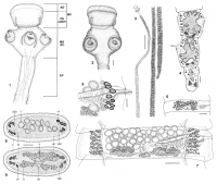
FIGURE 1. Notomegarhynchus navonae n. gen. et n. sp. Scolex terminology.
Abbreviations: AO, apical organ; CP, cephalic peduncle; MS, mesoscolex; MY, myzorhynchus; PS, proscolex. Terms used by Caira e... MoreFIGURE 1. Notomegarhynchus navonae n. gen. et n. sp. Scolex terminology.
Abbreviations: AO, apical organ; CP, cephalic peduncle; MS, mesoscolex; MY, myzorhynchus; PS, proscolex. Terms used by Caira et al. (1999) within parentheses: AMSP, apical modification of scolex proper; SP, scolex proper. FIGURES 2-7. Notomegarhynchus navonae n. gen. et n. sp. 2. Scolex, dorsoventral view, scale bar = 200 µm. 3. Complete worm, scale bar = 3 mm. 4. Terminal segments, scale bar = 200 µm. 5. Detail of terminal genitalia, scale bar = 100 µm. 6. Immature segment, scale bar = 200 µm. 7. Mature segment, scale bar = 200 µm. FIGURES 8, 9. Notomegarhynchus navonae n. gen. et n. sp. 8. Cross section of mature segment at level of testes anterior to cirrus sac. 9. Cross section of mature segment at level of ovarian isthmus. Scale bar = 200 µm. Abbreviations: C, cortex; DED, dorsal excretory duct; LM, longitudinal musculature; M, medulla; O, ovary; T, testis; V, vagina; VED, ventral excretory duct; VB vitelline follicles; UD, uteroduct. |
Line Drawing 2
|
Photo Micrograph
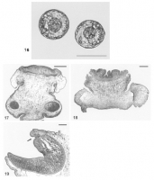
FIGURE 16. Notomegarhynchus navonae n. gen. et n. sp. Detail of eggs, scale bar = 50 µm. FIGURES 17-19. Histological sections. Figs. 17-19. Notomegarhynchus navonae n. gen. et n. sp. 17. Longitudinal ... MoreFIGURE 16. Notomegarhynchus navonae n. gen. et n. sp. Detail of eggs, scale bar = 50 µm. FIGURES 17-19. Histological sections. Figs. 17-19. Notomegarhynchus navonae n. gen. et n. sp. 17. Longitudinal section through a relaxed
scolex, scale bar = 400 µm. 18. Longitudinal section through a contracted scolex, scale bar = 200 µm. 19. Longitudinal section through
acetabulum, arrow indicaIes pit on anterior margin, scale bar = 100 µm. |
Scanning Electron Micrograph
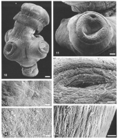
FIGURES 10-15. Scanning electron micrographs of Notomegarhynchus navonae n. gen. et n. sp. 10. Scolex, scale bar = 150 µm. 11. Detail of sucker-like acetabulum, scale bar = 60 µm. 12. Surface of myzor... MoreFIGURES 10-15. Scanning electron micrographs of Notomegarhynchus navonae n. gen. et n. sp. 10. Scolex, scale bar = 150 µm. 11. Detail of sucker-like acetabulum, scale bar = 60 µm. 12. Surface of myzorhynchus at level of apical organ, scale bar = 10 µm. 13. Detail of pit on anterior margin of acetabulum and distal acetabular surface, scale bar = 20 µm. 14. Interacetabular surface, scale bar = 10 µm. 15. Cephalic peduncle surface, scale bar = 10 µm. |
 FIGURE 1. Notomegarhynchus navonae n. gen. et n. sp. Scolex terminology.
Abbreviations: AO, apical organ; CP, cephalic peduncle; MS, mesoscolex; MY, myzorhynchus; PS, proscolex. Terms used by Caira et al. (1999) within parentheses: AMSP, apical modification of scolex proper; SP, scolex proper. FIGURES 2-7. Notomegarhynchus navonae n. gen. et n. sp. 2. Scolex, dorsoventral view, scale bar = 200 µm. 3. Complete worm, scale bar = 3 mm. 4. Terminal segments, scale bar = 200 µm. 5. Detail of terminal genitalia, scale bar = 100 µm. 6. Immature segment, scale bar = 200 µm. 7. Mature segment, scale bar = 200 µm. FIGURES 8, 9. Notomegarhynchus navonae n. gen. et n. sp. 8. Cross section of mature segment at level of testes anterior to cirrus sac. 9. Cross section of mature segment at level of ovarian isthmus. Scale bar = 200 µm. Abbreviations: C, cortex; DED, dorsal excretory duct; LM, longitudinal musculature; M, medulla; O, ovary; T, testis; V, vagina; VED, ventral excretory duct; VB vitelline follicles; UD, uteroduct.
FIGURE 1. Notomegarhynchus navonae n. gen. et n. sp. Scolex terminology.
Abbreviations: AO, apical organ; CP, cephalic peduncle; MS, mesoscolex; MY, myzorhynchus; PS, proscolex. Terms used by Caira et al. (1999) within parentheses: AMSP, apical modification of scolex proper; SP, scolex proper. FIGURES 2-7. Notomegarhynchus navonae n. gen. et n. sp. 2. Scolex, dorsoventral view, scale bar = 200 µm. 3. Complete worm, scale bar = 3 mm. 4. Terminal segments, scale bar = 200 µm. 5. Detail of terminal genitalia, scale bar = 100 µm. 6. Immature segment, scale bar = 200 µm. 7. Mature segment, scale bar = 200 µm. FIGURES 8, 9. Notomegarhynchus navonae n. gen. et n. sp. 8. Cross section of mature segment at level of testes anterior to cirrus sac. 9. Cross section of mature segment at level of ovarian isthmus. Scale bar = 200 µm. Abbreviations: C, cortex; DED, dorsal excretory duct; LM, longitudinal musculature; M, medulla; O, ovary; T, testis; V, vagina; VED, ventral excretory duct; VB vitelline follicles; UD, uteroduct.  FIGURE 16. Notomegarhynchus navonae n. gen. et n. sp. Detail of eggs, scale bar = 50 µm. FIGURES 17-19. Histological sections. Figs. 17-19. Notomegarhynchus navonae n. gen. et n. sp. 17. Longitudinal section through a relaxed
scolex, scale bar = 400 µm. 18. Longitudinal section through a contracted scolex, scale bar = 200 µm. 19. Longitudinal section through
acetabulum, arrow indicaIes pit on anterior margin, scale bar = 100 µm.
FIGURE 16. Notomegarhynchus navonae n. gen. et n. sp. Detail of eggs, scale bar = 50 µm. FIGURES 17-19. Histological sections. Figs. 17-19. Notomegarhynchus navonae n. gen. et n. sp. 17. Longitudinal section through a relaxed
scolex, scale bar = 400 µm. 18. Longitudinal section through a contracted scolex, scale bar = 200 µm. 19. Longitudinal section through
acetabulum, arrow indicaIes pit on anterior margin, scale bar = 100 µm.  FIGURES 10-15. Scanning electron micrographs of Notomegarhynchus navonae n. gen. et n. sp. 10. Scolex, scale bar = 150 µm. 11. Detail of sucker-like acetabulum, scale bar = 60 µm. 12. Surface of myzorhynchus at level of apical organ, scale bar = 10 µm. 13. Detail of pit on anterior margin of acetabulum and distal acetabular surface, scale bar = 20 µm. 14. Interacetabular surface, scale bar = 10 µm. 15. Cephalic peduncle surface, scale bar = 10 µm.

