Cestode Scientific Name
| Species ID | 3417 |
|---|---|
| Order | Rhinebothriidea |
| Family | Rhinebothriidae |
| Subfamily | |
| Genus | Rhinebothrium |
| Species | pearsoni |
| Authority | Butler, 1987 |
| Taxonomic Status | Valid |
| Valid Name | |
| Synonyms | |
| Genus Record | No |
| Type Species | No |
| Verified | Yes |
| Verified By | Reyda |
| Citation(s) |
Butler, S. A. 1987. Taxonomy of some tetraphyllidean cestodes from elasmobranch fishes. Australian Journal of Zoology 35: 343-371. (85) Download PDF |
| Redescription | |
| Scientific Name Notes |
Record Data
| Date (MM/DD/YYYY) | Action | User Name |
|---|---|---|
| 09/09/2006 | Created | K. Jensen , R. Tracy |
| 01/28/2015 | Modified | |
| 01/12/2016 | Modified | K. Mojica |
| 01/15/2016 | Modified | J. Caira |
| 01/21/2016 | Modified | K. Mojica |
Type Host
| Host Class | |||||||
|---|---|---|---|---|---|---|---|
| Host Order | Rhinopristiformes | ||||||
| Host Family | Rhinobatidae | ||||||
|
Type Host (Literal) |
|
||||||
|
Type Host (Valid) |
|
||||||
| Additional Host(s) | |||||||
| Site in Host | spiral intestine | ||||||
| Host Notes |
Type Locality
| Country | Australia |
|---|---|
| Body of Water | Moreton Bay |
| Island(s) | |
| City/Region | Queensland |
| Coordinates | |
| DD Latitude | |
| DD Longitude | |
| Additional Localities | |
| Locality Notes |
Specimens
| Type Material | QM GL4614 (holotype), QM GL4615-16 (paratypes) |
|---|---|
| Total Number of Type Specimens | 25 |
| Voucher Material | |
| Specimen Notes |
Data are given as in original description unless otherwise indicated.
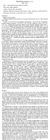


 FIGURES 1-6. Rhinebothrium pearsoni n. sp. 1. Scolex. 2. Detached mature segment, ventral view. 3. Everted cirrus, dorsal view. 4. Female reproductive system, ventral view. 5. Egg capsule. 6. Egg, containing hexacanth. Scale lines: 1, 0.4 mm; 2, 0.2 mm; 4, 5, 0.1 mm; 6, 30 µm.
  FIGURES 9-14. Rhinebothrium pearsoni n. sp. 9. Scolex, head-on view, note cephalic peduncle (arrowed). 10. Bothridium. 11. Scolex, lateral view, note pedicels (arrowed). 12. Junction of cephalic peduncle (arrowed) and beginning of segmentation, note microvilli on peduncle. 13. Attaching surface of bothridium, note short microvilli. 14. Surface of pedicel, note long microvilli. Scale lines: 9, 11. 200 µm. 10. 100µm. 12. 40 µm. 14. 10 µm. 13. 4 µm.
 Best viewed in Firefox


