Cestode Scientific Name
| Species ID | 3347 |
|---|---|
| Order | Phyllobothriidea |
| Family | |
| Subfamily | |
| Genus | Phyllobothrium |
| Species | vagans |
| Authority | Haswell, 1902 |
| Taxonomic Status | Incertae sedis |
| Valid Name | |
| Synonyms | |
| Genus Record | No |
| Type Species | No |
| Verified | No |
| Verified By | |
| Citation(s) |
Haswell, W. A. 1902. On a cestode from cestracion. Quarterly Journal of Microscopical Science 46: 399-415. (554) Download PDF |
| Redescription | |
| Scientific Name Notes | -Haswell (1902) did not use the scientific name for the host of Phyllobothrium vagans and no type locality is given; Williams (1968) indicates Haswells material was collected in New Zealand -Haswell (1902) begins with reference to the Port Jackson shark, but proposes the name P. vagans for the Cestracion parasite (pg. 401). |
Record Data
| Date (MM/DD/YYYY) | Action | User Name |
|---|---|---|
| 09/09/2006 | Created | T.R. Ruhnke and J.N. Caira |
| 02/05/2015 | Modified | |
| 06/06/2015 | Modified | J. Caira |
| 06/08/2015 | Modified | J. Caira |
| 06/13/2015 | Modified | J. Caira |
| 09/19/2020 | Modified | J. Caira |
Type Host
| Host Class | |||||||
|---|---|---|---|---|---|---|---|
| Host Order | Heterodontiformes | ||||||
| Host Family | Heterodontidae | ||||||
|
Type Host (Literal) |
|
||||||
|
Type Host (Valid) |
|
||||||
| Additional Host(s) | |||||||
| Site in Host | |||||||
| Host Notes |
Type Locality
| Country | |
|---|---|
| Body of Water | |
| Island(s) | |
| City/Region | |
| Coordinates | |
| DD Latitude | |
| DD Longitude | |
| Additional Localities | |
| Locality Notes |
Specimens
| Type Material | |
|---|---|
| Total Number of Type Specimens | |
| Voucher Material | |
| Specimen Notes |
Data are given as in original description unless otherwise indicated.
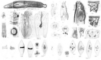

 PLATES 22-24. FIGURES 1-32. Phyllobothrium vagans n. sp. 1. Scolex magnified. 2. Last proglottis of strobila magnified. 3. Free proglottis, dorsal aspect. 4. Portion of transverse section of strobila. X600. 5. Transverse section of cirrus. cu. cuticle, with spinules. c.m. circular muscle. l.m. longitudinal muscle. my. myoblasts. 6. General view of female reproductive apparatus from ventral side. 7. Dorsal view of median part of ovary and ducts. 8. Section passing through swallowing apparatus, first part of oviduct and main vitelline duct X 450. 9. Section dorsal to that represented in Fig. 8, showing the vagina, recepticum seminis, and shell-gland X 450. 10. Section dorsal to that in Figure 9, showing recepticum seminis and fertilising duct X450. 11. Section passing through swallowing apparatus and median part of ovary. 12. Section immediately behind that in Fig 11. 13. Oblique series of mouth of swallowing apparatus X600. 14. Transverse series of swallowing apparatus and its relation to ovary. 15. Transverse series of swallowing apparatus to oviduct. 16. Transverse series of swallowing apparatus and oviduct X 600. 17. Horizontal series of oviduct where ducts of shell gland open into it; an ovum in the act of union with a yolk cell. 18. Transverse section of primary uterus, vagina, and ruptured secondary uterus. 19. Egg with unsegmented ovum. 20. Two-cell stage showing globules and concentrically laminated bodies of the vitelline mass. 21. Two-celled stage. 22. Three-celled stage. 23. Fouyr-celled stage. 24. Stage of about eight cells. 25. Surface view of blastoderm of somewhat later stage than fig 24. 26. Stage of about fourteen cells. 27. Surface view of disc-like blastoderm. 28-29. Disc-like stages seen edgewise. 30. Hexacanth embryo with the hooks retracted. 31. Hexacanth embryo with the hooks everted. 32. Egg containing hexacanth embryo. Fresh specimens.
 Best viewed in Firefox


