Line Drawing 1
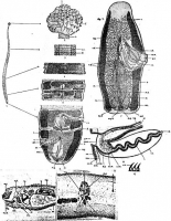
FIGURES 1-6, 7. Phyllobothrium sinuosiceps n. sp. 1. Complete strobila. 2. Anterior view. 3-6. Segments from different regions of the same complete strobila, dorsal view. 7. Free segment showing ... MoreFIGURES 1-6, 7. Phyllobothrium sinuosiceps n. sp. 1. Complete strobila. 2. Anterior view. 3-6. Segments from different regions of the same complete strobila, dorsal view. 7. Free segment showing male and female genitalia, dorsal view.
Abbreviations: b.m., basement membrane; c., cirrus; c.h., cirrus hooks; ci., cilia; c.s., cirrus sac; cu.m., cuticular membrane; d.l.m., deep longitudinal muscles; e.v., excretory vessel; f., folds of scolex; fl., posterior flap of segment; g.a., genital atrium; m.y.d., median yolk duct; m.g., Mehliss gland; n., neck; ov., ovary; ovc., ovicapt; ovd., oviduct; p.g., prostatic gland cell; r.s., receptaculum seminis; s.c., scaly cuticle; s.c.m., superficial circular muscles; s.l.m., superficial longitudinal muscles; t., testis; t.y.d., transverse yolk duct; ut., uterus; ut.d., uterine duct; v.d., vas deferens; v.ef., vas eferens; v.g., vagina; y., yolk gland. |
Line Drawing 2
|
Photo Micrograph
|
Scanning Electron Micrograph
|

 FIGURES 1-6, 7. Phyllobothrium sinuosiceps n. sp. 1. Complete strobila. 2. Anterior view. 3-6. Segments from different regions of the same complete strobila, dorsal view. 7. Free segment showing male and female genitalia, dorsal view.
Abbreviations: b.m., basement membrane; c., cirrus; c.h., cirrus hooks; ci., cilia; c.s., cirrus sac; cu.m., cuticular membrane; d.l.m., deep longitudinal muscles; e.v., excretory vessel; f., folds of scolex; fl., posterior flap of segment; g.a., genital atrium; m.y.d., median yolk duct; m.g., Mehliss gland; n., neck; ov., ovary; ovc., ovicapt; ovd., oviduct; p.g., prostatic gland cell; r.s., receptaculum seminis; s.c., scaly cuticle; s.c.m., superficial circular muscles; s.l.m., superficial longitudinal muscles; t., testis; t.y.d., transverse yolk duct; ut., uterus; ut.d., uterine duct; v.d., vas deferens; v.ef., vas eferens; v.g., vagina; y., yolk gland.
FIGURES 1-6, 7. Phyllobothrium sinuosiceps n. sp. 1. Complete strobila. 2. Anterior view. 3-6. Segments from different regions of the same complete strobila, dorsal view. 7. Free segment showing male and female genitalia, dorsal view.
Abbreviations: b.m., basement membrane; c., cirrus; c.h., cirrus hooks; ci., cilia; c.s., cirrus sac; cu.m., cuticular membrane; d.l.m., deep longitudinal muscles; e.v., excretory vessel; f., folds of scolex; fl., posterior flap of segment; g.a., genital atrium; m.y.d., median yolk duct; m.g., Mehliss gland; n., neck; ov., ovary; ovc., ovicapt; ovd., oviduct; p.g., prostatic gland cell; r.s., receptaculum seminis; s.c., scaly cuticle; s.c.m., superficial circular muscles; s.l.m., superficial longitudinal muscles; t., testis; t.y.d., transverse yolk duct; ut., uterus; ut.d., uterine duct; v.d., vas deferens; v.ef., vas eferens; v.g., vagina; y., yolk gland. 

