Line Drawing 1
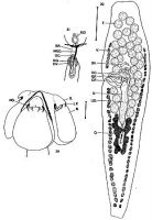
FIGURES 29-31. Pedibothrium ottleyi n. sp. 29. Scolex. 30. Mature segment, ventral view. 31. Female reproducive system, ventral view. Scale bars: 29 & 31, 0.1 mm; 30, 0.2 mm. Abbreviations: B., ... MoreFIGURES 29-31. Pedibothrium ottleyi n. sp. 29. Scolex. 30. Mature segment, ventral view. 31. Female reproducive system, ventral view. Scale bars: 29 & 31, 0.1 mm; 30, 0.2 mm. Abbreviations: B., bothridium; C.C., copulatory canal; C.P., cirrus pouch; G.P., genital pore; H.O., hook; L.P., lateral projection; M.V.D.; median vitelline duct; O., ovary; O.C., oocapt; O.O., ootype; O.V., oviduct; S., sucker; S.R., seminal receptacle; S.V., seminal vessicle; T., testis; U., uterus; U.D., uterine duct; V., vitellaria. |
Line Drawing 2
|
Photo Micrograph
|
Scanning Electron Micrograph
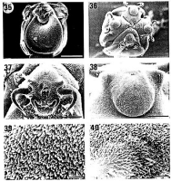
FIGURES 35-40. Pedibothrium ottleyi n. sp. 35. Scolex, lateral view. 36. Scolex, head-on view. 37. Hooks. 38. Accessory sucker, head-on view, note lateral projections. 39. Attaching surface of b... MoreFIGURES 35-40. Pedibothrium ottleyi n. sp. 35. Scolex, lateral view. 36. Scolex, head-on view. 37. Hooks. 38. Accessory sucker, head-on view, note lateral projections. 39. Attaching surface of bothridium, note finger-like processes. 40. Surface of accessory sucker, note hair-like processes. Scale bars: 35 & 36.,100 µm; 37, 40 µm; 38, 20 µm; 39 & 40, 4 µm. |
 FIGURES 29-31. Pedibothrium ottleyi n. sp. 29. Scolex. 30. Mature segment, ventral view. 31. Female reproducive system, ventral view. Scale bars: 29 & 31, 0.1 mm; 30, 0.2 mm. Abbreviations: B., bothridium; C.C., copulatory canal; C.P., cirrus pouch; G.P., genital pore; H.O., hook; L.P., lateral projection; M.V.D.; median vitelline duct; O., ovary; O.C., oocapt; O.O., ootype; O.V., oviduct; S., sucker; S.R., seminal receptacle; S.V., seminal vessicle; T., testis; U., uterus; U.D., uterine duct; V., vitellaria.
  FIGURES 35-40. Pedibothrium ottleyi n. sp. 35. Scolex, lateral view. 36. Scolex, head-on view. 37. Hooks. 38. Accessory sucker, head-on view, note lateral projections. 39. Attaching surface of bothridium, note finger-like processes. 40. Surface of accessory sucker, note hair-like processes. Scale bars: 35 & 36.,100 µm; 37, 40 µm; 38, 20 µm; 39 & 40, 4 µm.

