Cestode Scientific Name
| Species ID | 3241 |
|---|---|
| Order | Tetraphyllidea |
| Family | Balanobothriidae |
| Subfamily | |
| Genus | Pedibothrium |
| Species | globicephalum |
| Authority | Linton, 1908 |
| Taxonomic Status | Valid |
| Valid Name | |
| Synonyms | |
| Genus Record | No |
| Type Species | Yes |
| Verified | No |
| Verified By | |
| Citation(s) |
Linton, E. 1908. IX. Helminth fauna of the Dry Tortugas. I. Cestodes. Publication No. 102, Papers from the Tortugas Laboratory of the Carnegie Institution of Washington 1: 157-190. (277) Download PDF |
| Redescription |
Caira, J. N. 1992. Verification of multiple species of Pedibothrium in the Atlantic nurse shark with comments on the Australasian members of the genus. Journal of Parasitology 78: 289-308. (95) Download PDF |
| Scientific Name Notes | Caira & Pritchard (1996) used the date 1909 as the date the publication was distributed. However, this requires verification. |
Record Data
| Date (MM/DD/YYYY) | Action | User Name |
|---|---|---|
| 09/09/2006 | Created | K. Jensen , R. Tracy |
| 01/02/2014 | Modified | |
| 06/06/2015 | Modified | J. Caira |
| 06/08/2015 | Modified | J. Caira |
| 01/10/2016 | Modified | J. Caira |
| 01/13/2016 | Modified | J. Caira |
| 01/19/2016 | Modified | K. Jensen |
| 05/04/2016 | Modified | B. Barbeau |
| 09/18/2016 | Modified | K. Jensen |
| 08/07/2017 | Modified | J. Caira |
Type Host
| Host Class | |||||||
|---|---|---|---|---|---|---|---|
| Host Order | Orectolobiformes | ||||||
| Host Family | Ginglymostomatidae | ||||||
|
Type Host (Literal) |
|
||||||
|
Type Host (Valid) |
|
||||||
| Additional Host(s) | |||||||
| Site in Host | spiral intestine | ||||||
| Host Notes |
Type Locality
| Country | U.S.A. |
|---|---|
| Body of Water | |
| Island(s) | Dry Tortugas |
| City/Region | Florida |
| Coordinates | |
| DD Latitude | |
| DD Longitude | |
| Additional Localities | |
| Locality Notes |
Specimens
| Type Material | Holotype (USNM no. 8995); paratypes (3 specimens, USNM no. 8995); voucher specimens collected from G. cirratum in Bimini, Bahamas, May 1988 (10 specimens, USNM no. 82045; 10 specimens, HWML no. 34148; 2 slides of sections, HWML no. 34149; 5 specimens on stub for SEM, retained in collection of author). |
|---|---|
| Total Number of Type Specimens | 21 |
| Voucher Material | |
| Specimen Notes | Holotype (USNM no. 8995); paratypes (3 specimens, USNM no. 8995); voucher specimens collected from G. cirratum in Bimini, Bahamas, May 1988 (10 specimens, USNM no. 82045; 10 specimens, HWML no. 34148; 2 slides of sections, HWML no. 34149; 5 specimens on stub for SEM, retained in collection of author). |
Data are given as in original description unless otherwise indicated.

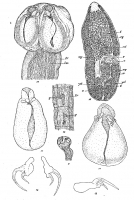
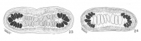
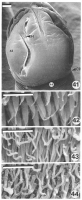
 PLATE 2. FIGURES 9-16. Pedibothrium globicephalum n. sp. 9. Head and neck, flattened, life; actual diameter of head 1.8 mm. 10. Head and neck, balsam; actual diameter of head 1 mm. 11, 12. Single bothria, life; actual lengths 0.86 and 0.92 mm. 13. Typical hook, side view; actual length 0.086 mm. 14. Pair of hooks, from small specimen; actual length 0.085 mm. 15. Segments with rudiments of reproductive organs, balsam; actual breadth 0.35 mm. 16. Free, mature segment, balsam; actual length 1.8 mm. Abbreviations: c, cirrus; cp, cirrus pouch; g, rudiment of genitalia; lm, longitudinal muscle; lv, lateral vessel; o, ovary; t, testes; u, uterus; v, vagina; vd, vas deferens; vg, vitelline glands; yd, vitelline duct.
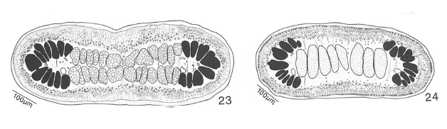 23. Section through ovarian region of voucher (HWML no. 34149) of P. globicephalum. 24. Section through region of testes of P. globicephalum. B, border between cortex and medulla; DE, dorsal
excretory duct; ED, excretory duct; NC, nerve cord; 0, ovary; T, testis; V, vitellaria; VD, vas deferens; VE,
ventral excretory duct.
23. Section through ovarian region of voucher (HWML no. 34149) of P. globicephalum. 24. Section through region of testes of P. globicephalum. B, border between cortex and medulla; DE, dorsal
excretory duct; ED, excretory duct; NC, nerve cord; 0, ovary; T, testis; V, vitellaria; VD, vas deferens; VE,
ventral excretory duct.  FIGURES 41-44. Scanning electron micrographs of Pedibothrium globicephalum. 41. Scolex; locations at which Figures 43, 44 were taken are indicated with
corresponding numbers; arrow at H marks location of hooks; note they are embedded in scolex musculature; arrow at CP marks end of cephalic peduncle. 42. Microtriches on distal bothridial surface, visible after mechanical
removal of outer bothridial margins. 43. Microtriches
on proximal bothridial surface. 44.
Microtriches on cephalic peduncle. Scale bar in Figure 41, 100 um; scale bar in Figures 42-44, 2 um
FIGURES 41-44. Scanning electron micrographs of Pedibothrium globicephalum. 41. Scolex; locations at which Figures 43, 44 were taken are indicated with
corresponding numbers; arrow at H marks location of hooks; note they are embedded in scolex musculature; arrow at CP marks end of cephalic peduncle. 42. Microtriches on distal bothridial surface, visible after mechanical
removal of outer bothridial margins. 43. Microtriches
on proximal bothridial surface. 44.
Microtriches on cephalic peduncle. Scale bar in Figure 41, 100 um; scale bar in Figures 42-44, 2 um Best viewed in Firefox


