Cestode Scientific Name
| Species ID | 3168 |
|---|---|
| Order | Phyllobothriidea |
| Family | Phyllobothriidae |
| Subfamily | |
| Genus | Flexibothrium |
| Species | ruhnkei |
| Authority | McKenzie & Caira, 1998 |
| Taxonomic Status | Valid |
| Valid Name | |
| Synonyms | |
| Genus Record | No |
| Type Species | Yes |
| Verified | No |
| Verified By | |
| Citation(s) |
McKenzie, V. J. and J. N. Caira. 1998. Three new genera and species of tapeworms from the longnose sawshark, Pristiophorus cirratus, with comments on their modes of attachment to the spiral intestine. Journal of Parasitology 84(2): 409-421. (306) Download PDF |
| Redescription | |
| Scientific Name Notes |
Record Data
| Date (MM/DD/YYYY) | Action | User Name |
|---|---|---|
| 09/09/2006 | Created | K. Jensen , R. Tracy |
| 02/19/2015 | Modified | |
| 06/06/2015 | Modified | T. Ruhnke |
| 10/09/2020 | Modified | B. Barbeau |
Type Host
| Host Class | |||||||
|---|---|---|---|---|---|---|---|
| Host Order | |||||||
| Host Family | |||||||
|
Type Host (Literal) |
|
||||||
|
Type Host (Valid) |
|
||||||
| Additional Host(s) | |||||||
| Site in Host | |||||||
| Host Notes |
Type Locality
| Country | |
|---|---|
| Body of Water | |
| Island(s) | |
| City/Region | |
| Coordinates | |
| DD Latitude | |
| DD Longitude | |
| Additional Localities | |
| Locality Notes |
Specimens
| Type Material | QM No. G214121 (holotype) QM No. G214122-G214129 AHC No. 27924-27929 USNPC No. 87577 HWML No. 39625 |
|---|---|
| Total Number of Type Specimens | 20 |
| Voucher Material | |
| Specimen Notes |
Data are given as in original description unless otherwise indicated.
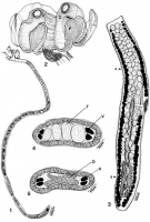

 FIGURES 1-5. Line drawings of Flexibothrium ruhnkei n. g., n. sp. 1. Entire worm. 2. Scolex. 3. Mature segment. 4. Cross section through mature segment at postition indicated by arrow numbered 4 in Figure 3. 5. Cross section through ovarian isthmus in mature segment at position indicated by arrow numbered 5 in Figure 3. D, distal bothridial surface; O, ovary; P, proximal borthridial surface; T, testis; V, vitellaria.
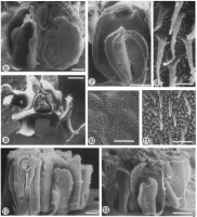
  FIGURE6S- 13. Scanning electron micrographs of Flexibothrium ruhnkei n. g., n. sp. 6. Scolex. 7. Enlarged view of bothridium. Location at which Figures 8, 10, 11 were taken are indicated with corresponding numbers. 8. Microtriches on proximal surface of flexed up portion of
bothridium. 9. Apical view of scolex with villus (V) retained in interbothridial space. Note open-faced grooves of bothridium (G), and flipped up portion of bothridium (F). 10. Microtriches on distal bothridial surface near margin of apical sucker. 11. Microtriches on face of distal bothridial surface. 12. Scolex attached to mucosal tissue. Note flipped up portions of 2 bothridia (F) making intimate contact with villi (V). 13. Scolex attached to mucosal tissue with villi removed from 2 open-faced grooves of 1 bothridium. Scale bar in Figures 6, 7, 9, 12, 100 µm; Figures 8,
10, 11, 1 µm; Figure 13, 200 µm.
FIGURE6S- 13. Scanning electron micrographs of Flexibothrium ruhnkei n. g., n. sp. 6. Scolex. 7. Enlarged view of bothridium. Location at which Figures 8, 10, 11 were taken are indicated with corresponding numbers. 8. Microtriches on proximal surface of flexed up portion of
bothridium. 9. Apical view of scolex with villus (V) retained in interbothridial space. Note open-faced grooves of bothridium (G), and flipped up portion of bothridium (F). 10. Microtriches on distal bothridial surface near margin of apical sucker. 11. Microtriches on face of distal bothridial surface. 12. Scolex attached to mucosal tissue. Note flipped up portions of 2 bothridia (F) making intimate contact with villi (V). 13. Scolex attached to mucosal tissue with villi removed from 2 open-faced grooves of 1 bothridium. Scale bar in Figures 6, 7, 9, 12, 100 µm; Figures 8,
10, 11, 1 µm; Figure 13, 200 µm. Best viewed in Firefox


