Line Drawing 1
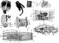
PLATE 1. FIGURES 2 & 3. Dinobothrium planum n. sp. 2. Dinobothrium planum, from Cetorhinus maximus. Front view of scolex. Length of bothrium 7mm. 3. Marginal view of scolex. Length of bothrium 7mm... MorePLATE 1. FIGURES 2 & 3. Dinobothrium planum n. sp. 2. Dinobothrium planum, from Cetorhinus maximus. Front view of scolex. Length of bothrium 7mm. 3. Marginal view of scolex. Length of bothrium 7mm.
PLATE 3. FIGURES 7-10. Dinobothrium planum n. sp. 7. Dinobothrium planum. A. Portion of strobile with ripe proglottides. Breadth 4mm. For explanation of lettering see p. 9. B. Eggs. Diameters, 0.012 to 0.015mm. 8. Dinobothrium plamun. A. Sagittal section near lateral margin of proglottis. Length of proglottis, 0.4 mm. For explanation of lettering see p. 9. B. Spines from cirrus. Length, 0.015mm. 9. Dinobothrium planum. Mediam sagittal section of proglottis, partly diagrammatic, more than one section having been used in representing the anatomy in the region of the shell gland. Length of proglottis, 0.4mm. For explanation of lettering see p. 9. 10. Dinobothrium planum. Transverse section of mature proglottis, partly diagrammatic. abouth three sections having been used in representing the vagina and cirrus. Length of longer diameter of section, 3mm. For explanation of lettering see p. 9.
PLATE 4. FIGURES 11-13. Dinobothrium planum n. sp. 11. Dinobothrium planum. Median sagittal section of immature proglottis, partly diagrammatic, more than one section having been used in representing the anatomy in the region of the shell gland. Length of proglottis, 0.4mm. For explanation of lettering see p. 9. 12. Dinobothrium planum. Diagram of part of female reproductive organs. For explanation of lettering see p. 9. 13. Stereogram of mature proglottis, view from ventral side. For explanation of lettering see p. 9. [Abbreviatitons:] c, cuticle; cp, cirrus pouch; ev, excretory vessel; evd, dorsal excretory vessel; evv, ventral excretory vessel; ga, genital aperture; gc, ganglion cells; gd, germ duct; lm, longitudinal muscles; n, nerve; o, ovary (germarium); ov, oviduct; sc, subcuticula; scg, subcuticular glands; sd, sperm duct; sg, shell-gland; sr, seminal receptacle; t, testes; u, uterusa; v, vagina; vd, vas deferens; vg, vitellaria; yd, yolk duct. |
Line Drawing 2
|
Photo Micrograph
|
Scanning Electron Micrograph
|
 PLATE 1. FIGURES 2 & 3. Dinobothrium planum n. sp. 2. Dinobothrium planum, from Cetorhinus maximus. Front view of scolex. Length of bothrium 7mm. 3. Marginal view of scolex. Length of bothrium 7mm.
PLATE 3. FIGURES 7-10. Dinobothrium planum n. sp. 7. Dinobothrium planum. A. Portion of strobile with ripe proglottides. Breadth 4mm. For explanation of lettering see p. 9. B. Eggs. Diameters, 0.012 to 0.015mm. 8. Dinobothrium plamun. A. Sagittal section near lateral margin of proglottis. Length of proglottis, 0.4 mm. For explanation of lettering see p. 9. B. Spines from cirrus. Length, 0.015mm. 9. Dinobothrium planum. Mediam sagittal section of proglottis, partly diagrammatic, more than one section having been used in representing the anatomy in the region of the shell gland. Length of proglottis, 0.4mm. For explanation of lettering see p. 9. 10. Dinobothrium planum. Transverse section of mature proglottis, partly diagrammatic. abouth three sections having been used in representing the vagina and cirrus. Length of longer diameter of section, 3mm. For explanation of lettering see p. 9.
PLATE 4. FIGURES 11-13. Dinobothrium planum n. sp. 11. Dinobothrium planum. Median sagittal section of immature proglottis, partly diagrammatic, more than one section having been used in representing the anatomy in the region of the shell gland. Length of proglottis, 0.4mm. For explanation of lettering see p. 9. 12. Dinobothrium planum. Diagram of part of female reproductive organs. For explanation of lettering see p. 9. 13. Stereogram of mature proglottis, view from ventral side. For explanation of lettering see p. 9. [Abbreviatitons:] c, cuticle; cp, cirrus pouch; ev, excretory vessel; evd, dorsal excretory vessel; evv, ventral excretory vessel; ga, genital aperture; gc, ganglion cells; gd, germ duct; lm, longitudinal muscles; n, nerve; o, ovary (germarium); ov, oviduct; sc, subcuticula; scg, subcuticular glands; sd, sperm duct; sg, shell-gland; sr, seminal receptacle; t, testes; u, uterusa; v, vagina; vd, vas deferens; vg, vitellaria; yd, yolk duct.
PLATE 1. FIGURES 2 & 3. Dinobothrium planum n. sp. 2. Dinobothrium planum, from Cetorhinus maximus. Front view of scolex. Length of bothrium 7mm. 3. Marginal view of scolex. Length of bothrium 7mm.
PLATE 3. FIGURES 7-10. Dinobothrium planum n. sp. 7. Dinobothrium planum. A. Portion of strobile with ripe proglottides. Breadth 4mm. For explanation of lettering see p. 9. B. Eggs. Diameters, 0.012 to 0.015mm. 8. Dinobothrium plamun. A. Sagittal section near lateral margin of proglottis. Length of proglottis, 0.4 mm. For explanation of lettering see p. 9. B. Spines from cirrus. Length, 0.015mm. 9. Dinobothrium planum. Mediam sagittal section of proglottis, partly diagrammatic, more than one section having been used in representing the anatomy in the region of the shell gland. Length of proglottis, 0.4mm. For explanation of lettering see p. 9. 10. Dinobothrium planum. Transverse section of mature proglottis, partly diagrammatic. abouth three sections having been used in representing the vagina and cirrus. Length of longer diameter of section, 3mm. For explanation of lettering see p. 9.
PLATE 4. FIGURES 11-13. Dinobothrium planum n. sp. 11. Dinobothrium planum. Median sagittal section of immature proglottis, partly diagrammatic, more than one section having been used in representing the anatomy in the region of the shell gland. Length of proglottis, 0.4mm. For explanation of lettering see p. 9. 12. Dinobothrium planum. Diagram of part of female reproductive organs. For explanation of lettering see p. 9. 13. Stereogram of mature proglottis, view from ventral side. For explanation of lettering see p. 9. [Abbreviatitons:] c, cuticle; cp, cirrus pouch; ev, excretory vessel; evd, dorsal excretory vessel; evv, ventral excretory vessel; ga, genital aperture; gc, ganglion cells; gd, germ duct; lm, longitudinal muscles; n, nerve; o, ovary (germarium); ov, oviduct; sc, subcuticula; scg, subcuticular glands; sd, sperm duct; sg, shell-gland; sr, seminal receptacle; t, testes; u, uterusa; v, vagina; vd, vas deferens; vg, vitellaria; yd, yolk duct. 

