Cestode Scientific Name
| Species ID | 3093 |
|---|---|
| Order | Tetraphyllidea |
| Family | |
| Subfamily | |
| Genus | Dinobothrium |
| Species | keilini |
| Authority | Sproston, 1948 |
| Taxonomic Status | Incertae sedis |
| Valid Name | |
| Synonyms | |
| Genus Record | No |
| Type Species | No |
| Verified | No |
| Verified By | |
| Citation(s) |
Sproston, N. G. 1948. On the genus Dinobothrium van Beneden (Cestoda), with a description of two new species from sharks, and a note on Monorygma sp. from the electric ray. Parasitology 39: 73-90. (452) Download PDF |
| Redescription | |
| Scientific Name Notes |
Record Data
| Date (MM/DD/YYYY) | Action | User Name |
|---|---|---|
| 09/09/2006 | Created | K. Jensen , R. Tracy |
| 02/18/2015 | Modified | |
| 06/06/2015 | Modified | J. Caira |
| 06/08/2015 | Modified | J. Caira |
| 01/13/2016 | Modified | K. Jensen |
| 05/19/2022 | Modified | J. Caira |
Type Host
| Host Class | |||||||
|---|---|---|---|---|---|---|---|
| Host Order | Carcharhiniformes | ||||||
| Host Family | Carcharhinidae | ||||||
|
Type Host (Literal) |
|
||||||
|
Type Host (Valid) |
|
||||||
| Additional Host(s) | |||||||
| Site in Host | spiral intestine | ||||||
| Host Notes |
Type Locality
| Country | Great Britain |
|---|---|
| Body of Water | |
| Island(s) | |
| City/Region | Plymouth |
| Coordinates | |
| DD Latitude | |
| DD Longitude | |
| Additional Localities | |
| Locality Notes |
Specimens
| Type Material | |
|---|---|
| Total Number of Type Specimens | 1 |
| Voucher Material | |
| Specimen Notes |
Data are given as in original description unless otherwise indicated.
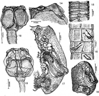
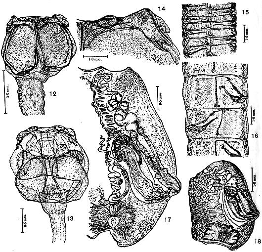 FIGURES 12-18. Dinobothrium keilini n. sp. 12. Scolex from mature strobilus (mounted half-scolex, seen from the outer face; owing to pressure the `bulbous vesicles behind the bothria are seen projecting on either side of them). 13. Scolex of very young strobilus (whole-mounted, nearer half slightly displaced downwards). 14. Bothridial crest (portion of half-scolex, mounted with pressure) showing true sucker and reflexed bifid lobe, here folded, at the outer end of crest. Direction of muscle fibres, as in other figures, shown by stippled lines. 15. Nascent proglottides (cf. Fig. 5). Note developing genital ducts and wide ventral cleft, here of different shape on the poral and aporal sides of the proglottis. The ventral excretory vessle in this species is throughout the strobilus much wider and more conspicuous than the dorsal; the inter- segmental openings of these vessles are never so obvious as in D. planum, and are not accompanied by furrows or notches in the body wall. 16. Young proglottides (cf. similar stage cin D. planum Fig. 6) The numerous testes are at an early stage but no vitellaria can be seen. Note the comparatively early development of the median region of the vagina in this and previous Fig., and yet the development of its distal portion lags far behind that of the cirrus-sac, which is more advanced than in Fig. 6. The fascia of longitudinal muscle fibers obliterated the ventral cleft from this dorsal view. 17. Dorsal view of portion of maturing proglottis at the beginning of the female phase: the vagina is distended with sperm and eggs are beginning to fill the uterine duct. Note the two sphincters on the vagina and the fine spines on its distal portion. 18. Dorsal view of mature, but not gravid, proglottis showing maximum developement of vitellaria, and ovary with medial shell-gland surrounding the oötype and fertilization chamber above; followed by the now very distended uterine duct, packed with developing eggs; the underlying uterus, destined to fill the median field, is as yet empty.
 Best viewed in Firefox


