Line Drawing 1
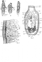
TEXT FIGURES 1-3. Biporophyllaeus madrassensis. n. sp. 1. a, b and c. Unmounted specimens. 2. Structure of body wall. 3. Diagramatic representation of the deposition of the various glands and du... MoreTEXT FIGURES 1-3. Biporophyllaeus madrassensis. n. sp. 1. a, b and c. Unmounted specimens. 2. Structure of body wall. 3. Diagramatic representation of the deposition of the various glands and ducts as viewed from the ventral side. Explanation of lettering: a. n. r., anterior nerve ring; b., bay; cu., cuticle; c. c., calcareous corpuscles; c. m., circular muscles; c. s., cirrus sac; e. d., excretory duct; e. v., excretory vesicle; f. c., fertilization chamber; fl. c., flame cell; g. a., gential atrium; g. c., gland cells; l. m., longitudinal chamber; fl. c., l. m. c., longitudinal muscle cells in probiscus; n., neck; n. c., nerve cord; n. g., posterior nerve ganglion; o. f., funnel of the oviduct; otp., ootype; ov., ovary; p., penis; pr., probiscis; p. n. r., posterior nerve ring; pr. n. r., probiscus nerve ring; s. c. l., subcuticular layer; s. d., duct of the shell gland; s. g., shell gland; sp., spinelets on the probiscus; tr., trunck; t. v., testicular vesicles; u., uterus; u. op., uterine opening; v., vagina; v. d., vas deferens; v. ev., valves of the excretory vesicle; v. f., valves of the oviducal funnel; v. g., vitelline glands; x., opening of uterus and vagina into the ootype. |
Line Drawing 2
|
Photo Micrograph
|
Scanning Electron Micrograph
|


 TEXT FIGURES 1-3. Biporophyllaeus madrassensis. n. sp. 1. a, b and c. Unmounted specimens. 2. Structure of body wall. 3. Diagramatic representation of the deposition of the various glands and ducts as viewed from the ventral side. Explanation of lettering: a. n. r., anterior nerve ring; b., bay; cu., cuticle; c. c., calcareous corpuscles; c. m., circular muscles; c. s., cirrus sac; e. d., excretory duct; e. v., excretory vesicle; f. c., fertilization chamber; fl. c., flame cell; g. a., gential atrium; g. c., gland cells; l. m., longitudinal chamber; fl. c., l. m. c., longitudinal muscle cells in probiscus; n., neck; n. c., nerve cord; n. g., posterior nerve ganglion; o. f., funnel of the oviduct; otp., ootype; ov., ovary; p., penis; pr., probiscis; p. n. r., posterior nerve ring; pr. n. r., probiscus nerve ring; s. c. l., subcuticular layer; s. d., duct of the shell gland; s. g., shell gland; sp., spinelets on the probiscus; tr., trunck; t. v., testicular vesicles; u., uterus; u. op., uterine opening; v., vagina; v. d., vas deferens; v. ev., valves of the excretory vesicle; v. f., valves of the oviducal funnel; v. g., vitelline glands; x., opening of uterus and vagina into the ootype.

