Cestode Scientific Name
| Species ID | 2831 |
|---|---|
| Order | Onchoproteocephalidea I |
| Family | Proteocephalidae |
| Subfamily | Proteocephalinae |
| Genus | Ophiotaenia |
| Species | alternans |
| Authority | Riser, 1942 |
| Taxonomic Status | Valid |
| Valid Name | |
| Synonyms | -Batrachotaenia alternans (Riser, 1942) Freze, 1965 -Proteocephalus alternans (Riser, 1942) Brooks, 1978 |
| Genus Record | No |
| Type Species | No |
| Verified | Yes |
| Verified By | adc |
| Citation(s) |
Riser, N. W. 1942. A new proteocephalid from Amphiuma tridactylum Cuvier. Transactions of the American Microscopical Society 61: 391-397. (4053) Download PDF |
| Redescription | |
| Scientific Name Notes | listed in Schmidt (1986) |
Record Data
| Date (MM/DD/YYYY) | Action | User Name |
|---|---|---|
| 03/21/2003 | Created | Scholz, Kuchta, Healy, Caira |
| 09/24/2006 | Modified | |
| 05/13/2016 | Modified | B. Barbeau |
| 02/06/2020 | Modified | B. Barbeau |
| 09/02/2020 | Modified | R. Kuchta |
| 12/03/2021 | Modified | B. Barbeau |
| 06/21/2024 | Modified | T. Scholz |
Type Host
| Host Class | Amphibia | ||||||
|---|---|---|---|---|---|---|---|
| Host Order | Urodela | ||||||
| Host Family | Amphiumidae | ||||||
|
Type Host (Literal) |
|
||||||
|
Type Host (Valid) |
|
||||||
| Additional Host(s) | |||||||
| Site in Host | small intestine | ||||||
| Host Notes |
Type Locality
| Country | U.S.A. |
|---|---|
| Body of Water | Reelfoot Lake |
| Island(s) | |
| City/Region | Tennessee |
| Coordinates | |
| DD Latitude | |
| DD Longitude | |
| Additional Localities | |
| Locality Notes | date collected: June 14, 1941 |
Specimens
| Type Material | USNM No. 36818 (type) (=USNPC) |
|---|---|
| Total Number of Type Specimens | |
| Voucher Material | |
| Specimen Notes |
Data are given as in original description unless otherwise indicated.


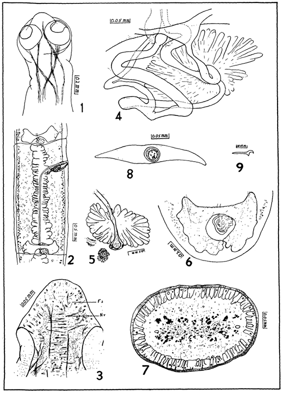 Fig. 1. Dorsal view of scolex. Fig. 2. Ventral view of gravid proglottid. Fig. 3. Longitudinal-diagonal section of scolex; nerve ring "Nr" and vestigial fifth sucker "Fs" showing. Fig. 4. Organs of the interovarian space. Fig. 5. Cross-section through oötype showing common vitelline duct and uterine passage in adjunction. Section of oviduct at lower left. Fig. 6. Posterior portion of an end proglottid showing characteristic shape of ovary. Fig. 7. Cross section through neck region. Muscle strands are solid black. Fig. 8. Egg as removed from living proglottid. Fig. 9. Onchosphere hook.
 Best viewed in Firefox


