Cestode Scientific Name
| Species ID | 2816 |
|---|---|
| Order | Onchoproteocephalidea I |
| Family | Proteocephalidae |
| Subfamily | Proteocephalinae |
| Genus | Macrobothriotaenia |
| Species | ficta |
| Authority | (Meggitt, 1931) Freze, 1965 |
| Taxonomic Status | Valid |
| Valid Name | |
| Synonyms | Crepidobothrium fictum Meggitt, 1931 Proteocephalus fictus (Meggitt, 1931) Huges, Baker & Dawson, 1941 |
| Genus Record | No |
| Type Species | Yes |
| Verified | No |
| Verified By | adc |
| Citation(s) |
Meggitt, F. J. 1931. On cestodes collected in Burma. Part II. . Parasitology 23: 250-263. (4035) Download PDFFreze, V. I. 1965. [Proteocephalata in Fish, Amphibians, and Reptiles] (In Russian). Osnovy Tsestodologii 5: 538 pp. (4031) Download PDF |
| Redescription |
Scholz, T., A. de Chambrier, R. Kuchta, D. T. J. Littlewood, and A. Waeschenbach. 2013. Macrobothriotaenia ficta (Cestoda: Proteocephalidea), a parasite of sunbeam snake (Xenopeltis unicolor): example of convergent evolution. Zootaxa 3640(3): 485-499. (6201) Download PDF |
| Scientific Name Notes | type of genus by designation. The date of the original species desription is cited by Freze (1965) incorrectly as 1927 |
Record Data
| Date (MM/DD/YYYY) | Action | User Name |
|---|---|---|
| 03/21/2003 | Created | Scholz, Kuchta, Healy, Caira |
| 02/16/2010 | Modified | |
| 12/18/2015 | Modified | N. Arisco |
| 06/30/2016 | Modified | B. Barbeau |
| 03/20/2018 | Modified | R. Kuchta |
| 03/15/2023 | Modified | R. Kuchta |
| 06/19/2024 | Modified | T. Scholz |
Type Host
| Host Class | Reptilia | ||||||
|---|---|---|---|---|---|---|---|
| Host Order | Squamata | ||||||
| Host Family | Xenopeltidae | ||||||
|
Type Host (Literal) |
|
||||||
|
Type Host (Valid) |
|
||||||
| Additional Host(s) | |||||||
| Site in Host | intestine | ||||||
| Host Notes |
Type Locality
| Country | Burma |
|---|---|
| Body of Water | |
| Island(s) | |
| City/Region | Rangoon (also Yangon) |
| Coordinates | |
| DD Latitude | |
| DD Longitude | |
| Additional Localities | |
| Locality Notes |
Specimens
| Type Material | - BMNH No. 1932.3.3.33 (type) |
|---|---|
| Total Number of Type Specimens | |
| Voucher Material | |
| Specimen Notes |
Data are given as in original description unless otherwise indicated.


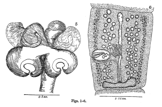
 Figures 11-16. Macrobothriotaenia ficta (Meggitt, 1931) from Xenopeltis unicolor. 11-13. Flattened scolex of mounted specimens (11, 12. adult specimen. 13. juvenile specimen, Vietnam, MHNG-PLAT 45476). 14, 15. Mature and pregravid proglottides, respectively, dorsal view, Thailand (BMNH 1976.4.13.14-15). 16. Gravid proglottis, ventral view, Vietnam (MHNG-PLAT 75475). Abbreviations: dc - dorsal osmoregulatory canals; vc - ventral osmoregulatory canals.
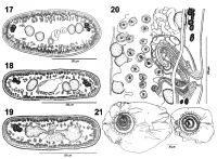
  Figures 17-21. Macrobothriotaenia ficta (Meggitt, 1931) from Xenopeltis unicolor. 17-19. Cross-sections of mature (17) and pregravid (18, 19) proglottides at level of anterior part (17, 18), and at level of ovary (19). 20. Terminal genitalia, ventral view, Thailand (BMNH 1976.4.13.14-15). 21. Eggs in distilled water, with collapsed outer hyaline envelope, Vietnam (MHNG-PLAT-75454). Abbreviations: dc-dorsal osmoregulatory canals; vc-ventral osmoregulatory canals.
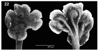
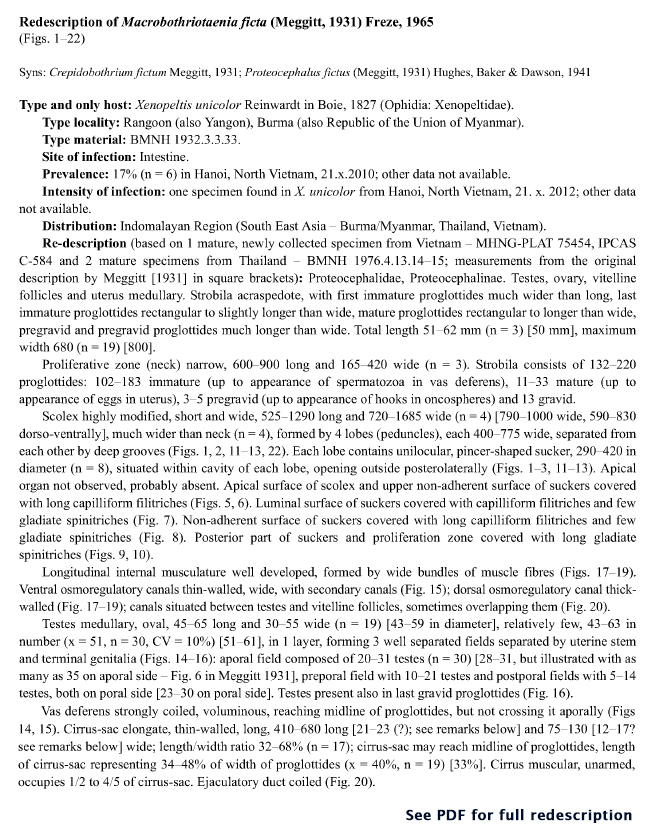
 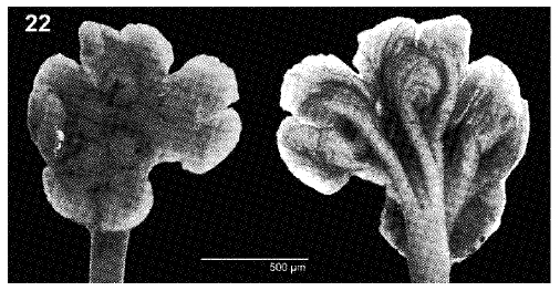 Figure 22. Macrobothriotaenia (Meggitt, 1931) from Xenopeltis unicolor. Unmounted scolex, apical view (left) and view from lower side (right), Vietnam (MHNG-PLAT 75454).
 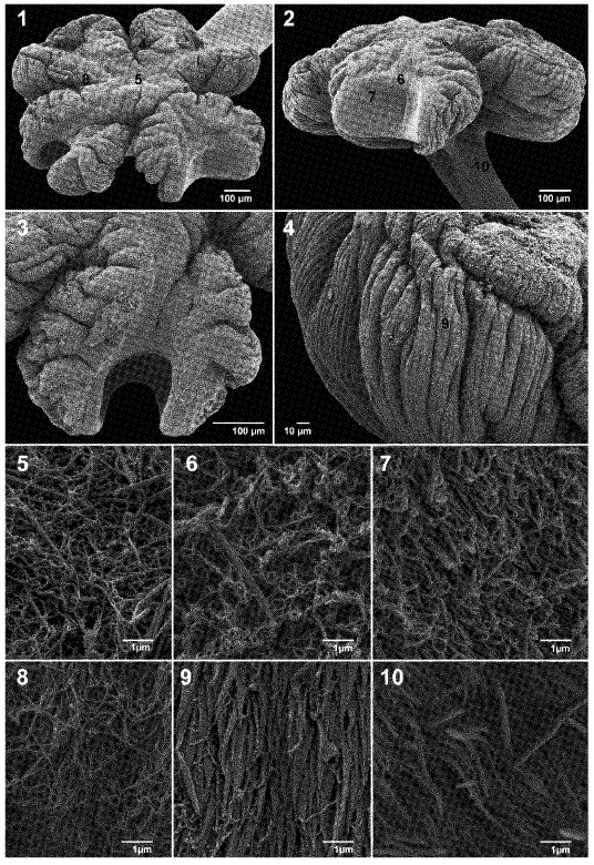 Figures 1-10. Macrobothriotaenia ficta (Meggitt, 1931) from Xenopeltis unicolor. Scanning electron micrographs. 1. Scolex, apical view. 2. Scolex, lateral view. 3. Detail of a sucker, apical view. 4. Lower part of the exterior surface of a sucker. 5. Capilliform filitriches at level of the apex surface. 6. Capilliform filitriches at the upper marginal surface of a sucker. 7. Luminal surface of suckers covered with capilliform filitriches and few gladiate spinitriches. 8. Non-adherent surface of suckers covered with long capilliform filitriches and few gladiate spinitriches. 9, 10. Posterior part of suckers and proliferation zone covered with long gladiate spinitriches. Note: small black numbers correspond to the figures showing higher magnification images of these surfaces. Figure 22. Macrobothriotaenia ficta (Meggit, 1931) from Xenopeltis unicolor. Unmounted scolex, apical view (left) and view from lower side (right), Vietnam (MHNG-PLAT 75454).
Best viewed in Firefox


