Line Drawing 1
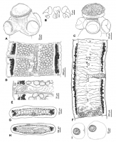
Figure 6. Gangesia macrones Woodland, 1924 from Sperata seenghala, India. A. Scolex, dorsoventral view (IPCAS C-618, field no. MS 4h). . Rostellar hooks (IPCAS C-618, field no. MS 4h). C. Scolex of de... MoreFigure 6. Gangesia macrones Woodland, 1924 from Sperata seenghala, India. A. Scolex, dorsoventral view (IPCAS C-618, field no. MS 4h). . Rostellar hooks (IPCAS C-618, field no. MS 4h). C. Scolex of decomposed specimen fixed with cold formalin and detached hooks and hooklets (IPCAS C-618, field no. MH 26). D. Mature proglottis, ventral view (MHNG-PLAT 82302, field no. MS 6r). E. Terminal genitalia, ventral view (IPCAS C-618, MS 4h). F. Gravid proglottis, ventral view (IPCAS C-618, field no. MS 4h). I. Eggs drawn in distilled water. G, H. Cross sections at level of testicular field and ovary, respectively (IPCAS C-618, field no. MS 4k). |
Line Drawing 2
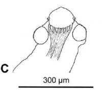
Figure 9. Outline of scoleces with retractor muscles of selected Gangesia taxa. C. G. macrones. |
Photo Micrograph
|
Scanning Electron Micrograph
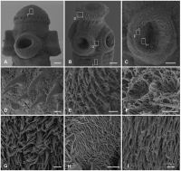
Figure 7. Scanning electron photomicrographs of the scolex of Gangesia macrones Woodland, 1924. A, B. Scolex, dorsoventral view. C. Sucker with small hooklets on the outer rim. D. Detail of a rostellu... MoreFigure 7. Scanning electron photomicrographs of the scolex of Gangesia macrones Woodland, 1924. A, B. Scolex, dorsoventral view. C. Sucker with small hooklets on the outer rim. D. Detail of a rostellum0like organ; note two rows of rostellar hooks of different shape with capilliform filitriches. E. Detail of the outer rim of the sucker with hooklets and capilliform filitriches. F. Detail of capilliform filitriches on the rostellum-like organ. G. Detail of capilliform filitriches and gladiate spinitriches in between the suckers. H. Detail of capilliform filitriches on the sucker. I. Gladiate spinitriches on the neck region. Scale bars: A, B -30 µm; C -20 µm; D-F -2 µm; G-I -1 µm. |
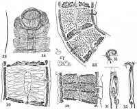
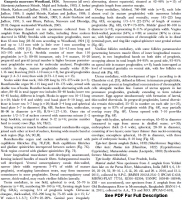

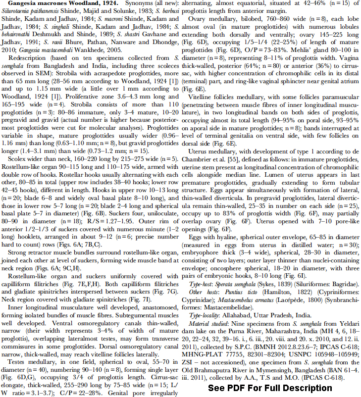
 Figure 6. Gangesia macrones Woodland, 1924 from Sperata seenghala, India. A. Scolex, dorsoventral view (IPCAS C-618, field no. MS 4h). . Rostellar hooks (IPCAS C-618, field no. MS 4h). C. Scolex of decomposed specimen fixed with cold formalin and detached hooks and hooklets (IPCAS C-618, field no. MH 26). D. Mature proglottis, ventral view (MHNG-PLAT 82302, field no. MS 6r). E. Terminal genitalia, ventral view (IPCAS C-618, MS 4h). F. Gravid proglottis, ventral view (IPCAS C-618, field no. MS 4h). I. Eggs drawn in distilled water. G, H. Cross sections at level of testicular field and ovary, respectively (IPCAS C-618, field no. MS 4k).
  Figure 9. Outline of scoleces with retractor muscles of selected Gangesia taxa. C. G. macrones.
  Figure 7. Scanning electron photomicrographs of the scolex of Gangesia macrones Woodland, 1924. A, B. Scolex, dorsoventral view. C. Sucker with small hooklets on the outer rim. D. Detail of a rostellum0like organ; note two rows of rostellar hooks of different shape with capilliform filitriches. E. Detail of the outer rim of the sucker with hooklets and capilliform filitriches. F. Detail of capilliform filitriches on the rostellum-like organ. G. Detail of capilliform filitriches and gladiate spinitriches in between the suckers. H. Detail of capilliform filitriches on the sucker. I. Gladiate spinitriches on the neck region. Scale bars: A, B -30 µm; C -20 µm; D-F -2 µm; G-I -1 µm.

