Cestode Scientific Name
| Species ID | 2748 |
|---|---|
| Order | Onchoproteocephalidea I |
| Family | Proteocephalidae |
| Subfamily | Gangesiinae |
| Genus | Gangesia |
| Species | bengalensis |
| Authority | (Southwell, 1913) Meggitt, 1927 |
| Taxonomic Status | Valid |
| Valid Name | |
| Synonyms | Ophryocotyle bengalensis Southwell, 1913 |
| Genus Record | No |
| Type Species | No |
| Verified | Yes |
| Verified By | T. Scholz |
| Citation(s) |
Southwell, T. 1913. Notes from the bengal Fisheries Laboratory, Indian Museum, No. 1. Records of the Indian Museum (a Journal of Indian Zoology) 9: 79-103 & plates . (4164) Download PDFMeggitt, F. J. 1927. Remarks on the cestode families Monticellidae and Ichthyotaeniidae. Annals of Tropical Medicine and Parasitology 21: 69-87. (4037) Download PDF |
| Redescription |
Ash, A., T. Scholz, A. de Chambrier, J. Brabec, M. Oros, P. K. Kar, S. P. Chavan, and J. Mariaux. 2012. Revision of Gangesia (Cestoda: Proteocephalidea) in the Indomalayan region: morphology, molecules and surface ultrastructure. PLOS One 7(10): e46421. (5987) Download PDF |
| Scientific Name Notes | -Meggitt (1927) does not explicitly state that this is a new combination, but this is the earliest use of this name we have found. -Meggitt (1927) pg. 74, listed Gangesia bengalensis (Sourthwell, 1922) as the type species of Gangesia Woodland, 1924, however, the correct type species is Gangesia wallago Woodland, 1924 |
Record Data
| Date (MM/DD/YYYY) | Action | User Name |
|---|---|---|
| 03/21/2003 | Created | A. Ash, Scholz, Kuchta, Healy, C |
| 10/25/2012 | Modified | |
| 10/15/2015 | Modified | N. Arisco |
| 10/21/2015 | Modified | N. Arisco |
| 04/22/2020 | Modified | T. Scholz |
| 04/24/2020 | Modified | T. Scholz |
| 12/03/2021 | Modified | B. Barbeau |
| 06/16/2024 | Modified | T. Scholz |
Type Host
| Host Class | Actinopterygii | ||||||
|---|---|---|---|---|---|---|---|
| Host Order | Cypriniformes | ||||||
| Host Family | Cyprinidae | ||||||
|
Type Host (Literal) |
|
||||||
|
Type Host (Valid) |
|
||||||
| Additional Host(s) | Wallago attu | ||||||
| Site in Host | |||||||
| Host Notes |
Type Locality
| Country | India |
|---|---|
| Body of Water | |
| Island(s) | |
| City/Region | Berhampur, West Bengal, India |
| Coordinates | |
| DD Latitude | |
| DD Longitude | |
| Additional Localities | |
| Locality Notes |
Specimens
| Type Material | -Indian Museum |
|---|---|
| Total Number of Type Specimens | |
| Voucher Material | |
| Specimen Notes |
Data are given as in original description unless otherwise indicated.
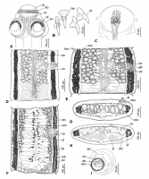

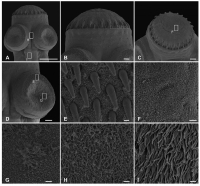
 Figure 1. Gangesia bengalensis (Southwell, 1913) from Wallago attu, India. A. Scolex, dorsoventral view (MHNG-PLAT 82308, field no. AA 133B). B. Rostellar hooks (IPCAS C-616, field no. AA 133). C. Detail of retractor muscles. D, E. Mature proglottides, ventral view (IPCAS C-616, field no. AA 133 and MHNG-PLAT 60721, field no. 147/08). F. Gravid proglottis, ventral view (IPCAS C-616, field no. AA 133). G, H. Cross sections at level of testicular field and ovary, respectively (IPCAS C-616, field no. AA 133); note that subtegumental layer is not fully illustrated. I. Egg drawn in distilled water. Abbreviations: ba - base of hook, bl - blade of hook, cs - cirrus-sac, doc - dorsal osmoregulatory canal, eh - embryonic hook, em - embryophore, ga - genital atrium, hl - hooklets, ho - hooks, ilm - internal longitudinal muscles, lub- lateral uterine branch, oe - outer envelope, on - oncosphere, ov - ovary, re - retractor muscles, ro - rostellum-like organ, sl - subtegumental layer, su - sucker, te - testes, us - uterine stem, uso - uterine slit-like opening, ut - uterus, va - vagina, vf - vitelline follicles, voc - ventral osmoregulatory canal, vs - vaginal sphincter.
 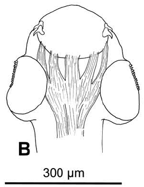 Figure 9. Outline of scoleces with retractor muscles of selected Gangesia taxa. B. Outline of the scoleces with retractor muscles of Gangesia agraensis.
 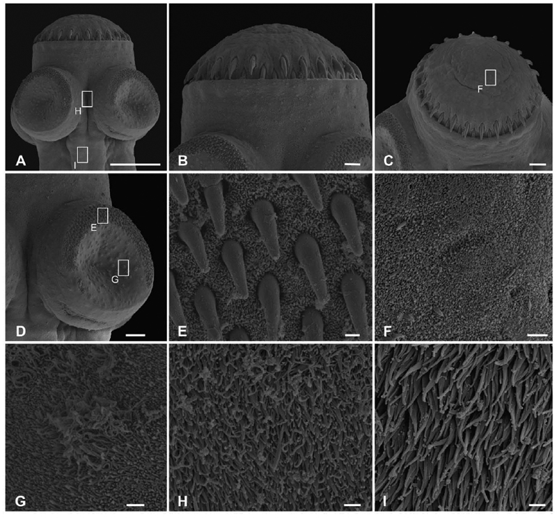 Figure 2. Scanning electron photomicrographs of Gangesia bengalensis (Southwell 1913). A. Scolex, dorsovenral view. B, C. Detail of rostellum-like organ; note two rows of rostellar hooks. D. Sucker with small hooklets on the outer rim. E. Detail of the outer rim of the sucker with hooklets. F. Detail of short, dense acicular filitriches on the rostellum-like organ. G. Detail of acicular filitriches and few capilliform filitriches on the sucker. H, Detail of capilliform filitriches and small gladiate spinitriches in between the suckers. I. Detail of gladiate spinitriches on the neck region. Scale bars: A -100 µm; B -10 µm; C, D -20 µm; E-I -µm.
Best viewed in Firefox


