Line Drawing 1
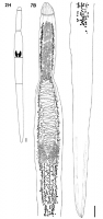
Fig. 2. Outlines of the body and individual body regions of Wenyonia spp. (ovary indicated in black; caudal portion separated by a dashed line): H, W. youdeoweii. Scale-bars: 1 mm. Fig. 7B, W. youdeow... MoreFig. 2. Outlines of the body and individual body regions of Wenyonia spp. (ovary indicated in black; caudal portion separated by a dashed line): H, W. youdeoweii. Scale-bars: 1 mm. Fig. 7B, W. youdeoweii Ukoli, 1972 from S. serrata, River Nile, Sudan. Entire worm, ventral (A) and dorsal (B) views. Scale-bars: 1mm. |
Line Drawing 2
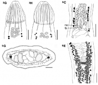
Fig. 3 G, H, W. youdeoweii Ukoli, 1972. Scale-bars: 500µm. Fig. 5 Cross-sections of Wenyonia spp. in the testicular (G) region. G, W. youdeoweii Ukoli, 1972. Scale-bars: G, 200µm. Fig. 9 C, E, W. youd... MoreFig. 3 G, H, W. youdeoweii Ukoli, 1972. Scale-bars: 500µm. Fig. 5 Cross-sections of Wenyonia spp. in the testicular (G) region. G, W. youdeoweii Ukoli, 1972. Scale-bars: G, 200µm. Fig. 9 C, E, W. youdeoweii Ukoli, 1972 from S. schall, River Nile. Cirrus-sac, ventral view. Abbreviations: mp, male genital pore; fp, female genital pore. Scale-bars: C,E, 500µm. |
Photo Micrograph
|
Scanning Electron Micrograph

Fig. 4 Scanning electron micrographs of M, N, W. youdeoweii Ukoli, 1972. Scale-bars: M, N, 300µm. |
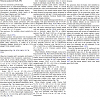
 Fig. 2. Outlines of the body and individual body regions of Wenyonia spp. (ovary indicated in black; caudal portion separated by a dashed line): H, W. youdeoweii. Scale-bars: 1 mm. Fig. 7B, W. youdeoweii Ukoli, 1972 from S. serrata, River Nile, Sudan. Entire worm, ventral (A) and dorsal (B) views. Scale-bars: 1mm.
 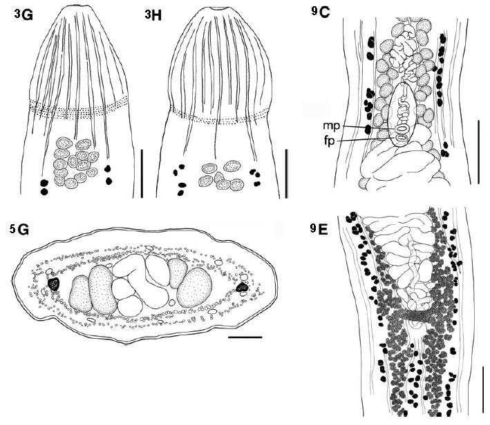 Fig. 3 G, H, W. youdeoweii Ukoli, 1972. Scale-bars: 500µm. Fig. 5 Cross-sections of Wenyonia spp. in the testicular (G) region. G, W. youdeoweii Ukoli, 1972. Scale-bars: G, 200µm. Fig. 9 C, E, W. youdeoweii Ukoli, 1972 from S. schall, River Nile. Cirrus-sac, ventral view. Abbreviations: mp, male genital pore; fp, female genital pore. Scale-bars: C,E, 500µm.
 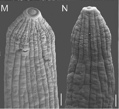 Fig. 4 Scanning electron micrographs of M, N, W. youdeoweii Ukoli, 1972. Scale-bars: M, N, 300µm.

