Cestode Scientific Name
| Species ID | 2562 |
|---|---|
| Order | Onchoproteocephalidea I |
| Family | Proteocephalidae |
| Subfamily | |
| Genus | Houssayela |
| Species | sudobim |
| Authority | (Woodland, 1935) Rego, 1987 |
| Taxonomic Status | Valid |
| Valid Name | |
| Synonyms | Myzophorus sudobim Woodland, 1935; Nomimoscolex woodlandi Freze, 1965 nec N. woodlandi Rego & Pavanelli, 1992] |
| Genus Record | No |
| Type Species | Yes |
| Verified | No |
| Verified By | adc |
| Citation(s) |
Woodland, W. N. F. 1935. Additional cestodes form the Amazon siluroids pirarara, dorad, and subobim. Proceedings of the Zoological Society of London Part 4: 851-862 +Plates . (3964) Download PDFRego, A. A. 1987. Cestoides proteocefalideos do Brasil. Reorganizacao taxonomica. Revista Brasileira de Biologia 47(1/2): 203-212. (3944) Download PDF |
| Redescription |
de Chambrier, A. and T. Scholz. 2005. Redescription of Houssayela sudobim (Woodland, 1935) (Cestoda: Proteocephalidea), a parasite of Pseudoplatystoma fasciatum (Pisces: Siluriformes) from the River Amazon. Systematic Parasitology 62: 161-169. (6638) Download PDF |
| Scientific Name Notes | Rego, 1987 misspelled sudobim as subodim Redescribed by de Chambrier & Scholz, 2005 |
Record Data
| Date (MM/DD/YYYY) | Action | User Name |
|---|---|---|
| 03/17/2003 | Created | Scholz, Kuchta, Healy, Caira |
| 02/16/2010 | Modified | |
| 12/03/2015 | Modified | N. Arisco |
| 12/06/2018 | Modified | P. Alves |
| 02/03/2020 | Modified | B. Barbeau |
| 12/03/2021 | Modified | B. Barbeau |
Type Host
| Host Class | |||||||
|---|---|---|---|---|---|---|---|
| Host Order | Siluriformes | ||||||
| Host Family | Pimelodidae | ||||||
|
Type Host (Literal) |
|
||||||
|
Type Host (Valid) |
|
||||||
| Additional Host(s) | |||||||
| Site in Host | |||||||
| Host Notes |
Type Locality
| Country | Brazil |
|---|---|
| Body of Water | Amazon River |
| Island(s) | |
| City/Region | Near Manaus and Santarém, Amazonas State |
| Coordinates | |
| DD Latitude | |
| DD Longitude | |
| Additional Localities | |
| Locality Notes |
Specimens
| Type Material | |
|---|---|
| Total Number of Type Specimens | |
| Voucher Material | |
| Specimen Notes |
Data are given as in original description unless otherwise indicated.

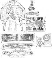


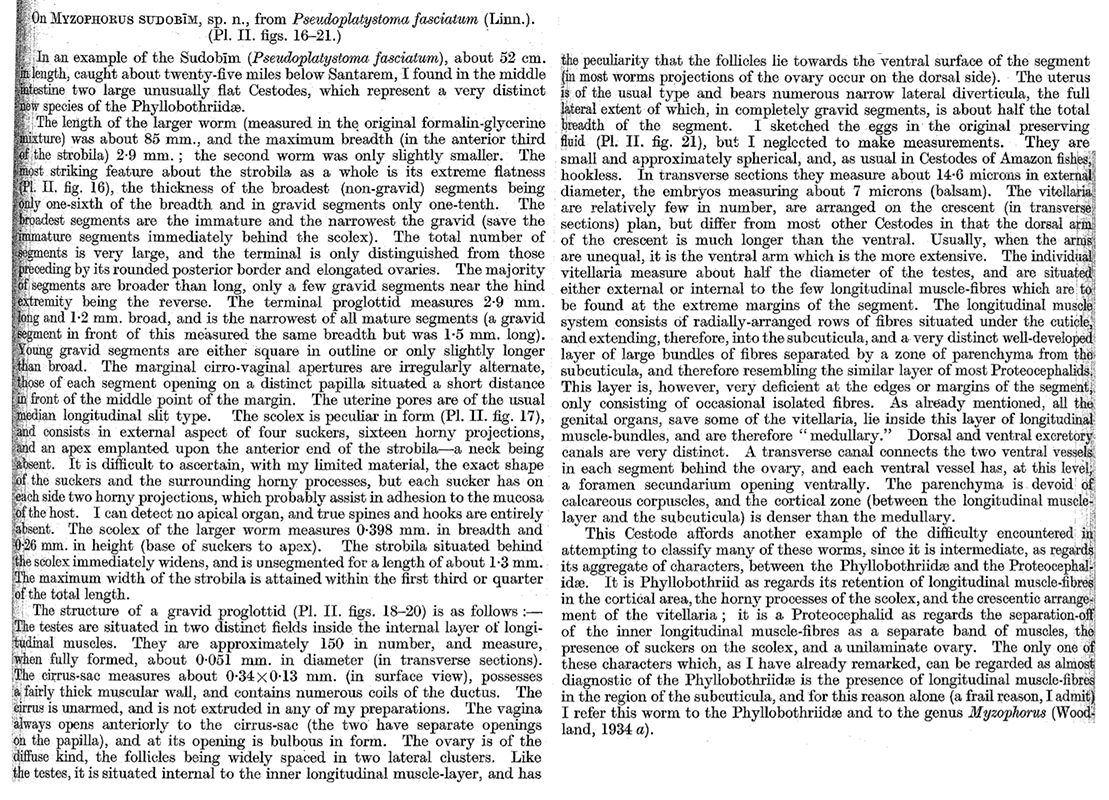
 Plate II. Myzophorus sudobim, sp. n. (Figs. 16-21.) Fig. 16 (nat. size): the entire worm. 17 (x87): the scolex in outline. 18 (x12): a gravid proglottid (ventral aspect). 19 (x39): portion of transverse section of mature proglottid anterior to ovary. 20 (x56): portion of transverse section of mature proglottid in region of ovary. 21 (x395): intra-uterine egg sketched in formalin. Lettering to Figures. CS. Cirrus-sac. DV. Dorsal excretory canal. LM. Longitudinal muscles. LMB. Longitudinal muscle-band. O. Ovary. S. Sucker. SB. Limit of subcuticula layer. UT. Uterus. V. Vagina. VIT. Vitellaria. VS. Coils of vas deferens. VV. Ventral excretory canal.
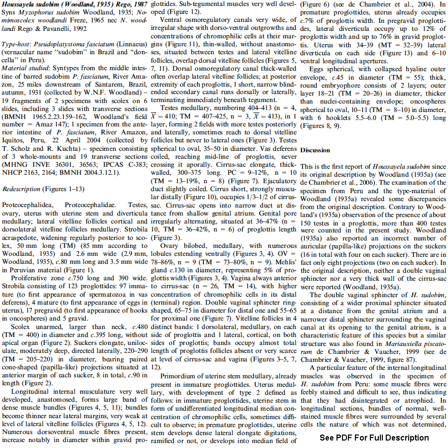
 Figure 2. Houssayela sudobim Woodland, 1935, IPCAS 383. Scolex, dorsoventral view, shwing the cone-shaped (papilla-like) projections of the sucker (arrow). Abbreviations: cg, cells with finely granular cytoplasm; do, dorsal osmoregulatroy canal; vo, ventral osmoregulatory canal. Scale-bar: 2, 250 µm. Figures 3-5. Houssayela sudobim Woodland, 1935. 3. Mature proglottis, ventral view, IPCAS 383. 4. Transverse section at the level of the ovary, INVE 36301. 5. Transverse section at the level of the middle of the proglottis and posterior to the cirrus-sac, showing the location of the future uterine aperture, INVE 36301. Figures 4 and 5 are oriented with the dorsal side uppermost. Abbreviations: do, dorsal osmoregulatory canal; lm, internal longitudinal musculature; ln, longitudinal lateral nerve; mg, Mehlis' gland; ov, ovary; te, testes; up, uterine pore; ut, uterus; vc, vaginal canal; vi, vitelline follicles; vo, ventral osmoregulatroy canal. Scale-bar: 3-5, 1,000 µm. Figures 6-9. Houssayela sudobim Woodland, 1935. 6. INVE 36301. Diagrammatic representation of type 2 progressive development of the uterus (see de Chambrier et al., 2004). 7. INVE 36301. Cirrus-sac and vagina, showing the double vaginal sphincter. 8. BMNH 1965.2.23.159-162 syntype material. Egg. 9. INVE 36301. Egg, Peruvian material. Abbrevations: cp, cirrus-sac; do, dorsal osmoregulatory canal; em, embryophore; mc, muscular part of the cirrus; oe, outer envelope; om, oncospheral membrane; on, oncosphere; va, vagina; vi, vitelline follicles; vo, ventral osmoregulatory canal; vs, vaginal sphincter. Scale-bars: 7, 500 µm; 8,9, 20 µm; 10, 100 µm.
 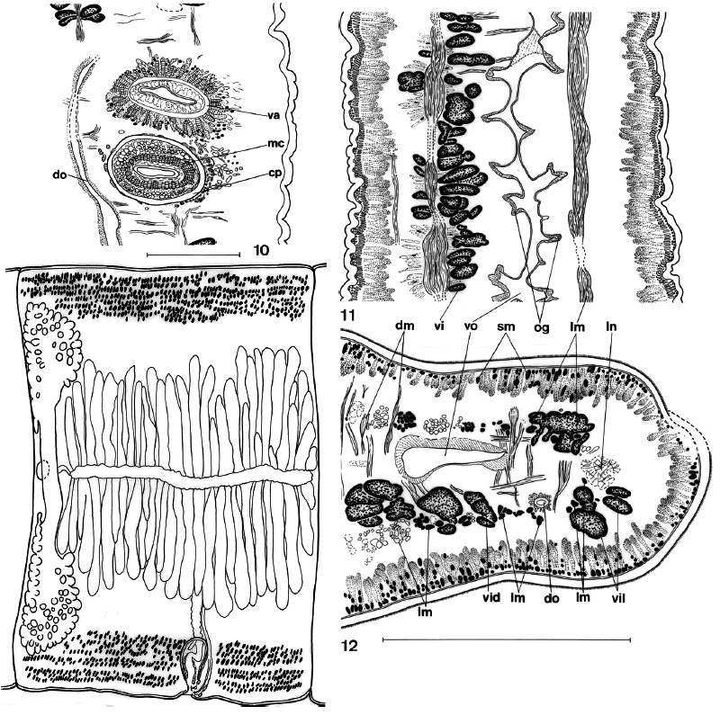 Figure 10. Houssayela sudobim Woodland, 1935. 10. INVE 36301. Sagittal section of the cirrus-sac and vagina at the level of the proximal vaginal sphincter. Abbrevations: cp, cirrus-sac; do, dorsal osmoregulatory canal; em, embryophore; mc, muscular part of the cirrus; oe, outer envelope; om, oncospheral membrane; on, oncosphere; va, vagina; vi, vitelline follicles; vo, ventral osmoregulatory canal; vs, vaginal sphincter. Scale-bars: 7, 500 µm; 8,9, 20 µm; 10, 100 µm. Figures 11-12. Houssayela sudobim Woodland, 1935. 11. INVE 36301. Sagittal section showing the irregular longitudinal internal musculature and outgrowths of the ventral osmoregulatory canal. 12. INVE 36301. Transverse section at the level of vitelline follicles; detail showing the cortical position of the lateral vitelline follicles and the medullary position of the dorsal follicles. The figure is oriented with the ventral surface uppermost. Abbreviations: dm, dorsoventral musculature; do, dorsal osmoregulatory canal; lm, internal longitudinal musculature; ln, longitudinals lateral nerve; og, outgrowths of the ventral osmoregulatory canal; sm, subtegumental musculature; vid, dorsal vitelline follicles; vil, lateral vitelline follicles; vi, vitelline follicles; vo, ventral osmoregulatory canal. Scale-bars: 11, 12, 250 µm. Figure 13. Houssayela sudobim Woodland, 1935. INVE 36801. Diagrammatic drawing showing the shape of the uterus in gravid proglottides, ventral view.
 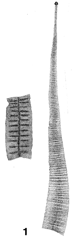 Figure 1. Houssayela sudobim Woodland, 1935, IPCAS 383. 1. Anterior part of the worm, diagrammatic.
Best viewed in Firefox


