Line Drawing 1

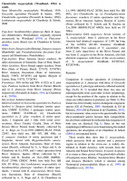
From Alves, etal 2017 (Cit# 7075). Fig. 3 Chambriella megacephala (Woodland, 1934) ex Zungaro jahu. A, Scolex, apical view (paratype of C. agostinhoi, CHIOC 32820c); B, Terminal genitalia, ventral vie... MoreFrom Alves, etal 2017 (Cit# 7075). Fig. 3 Chambriella megacephala (Woodland, 1934) ex Zungaro jahu. A, Scolex, apical view (paratype of C. agostinhoi, CHIOC 32820c); B, Terminal genitalia, ventral view (holotype of C. agostinhoi, CHIOC 32820a); C, Terminal genitalia, dorsal view (MHNH- PLAT 22025); D, E, Cross-sections at anterior and cirrus-sac level of proglottides, respectively (MHNH-PLAT 19546); F, Eggs drawn in distilled water (MHNH-PLAT 20768). Abbreviations: al, anterior sucker loculus; ci, cirrus; cs, cirrus-sac; do, dorsal osmoregulatory canal; ed, ejaculatory duct; em, bi-layered embryophore; lm, internal longitudinal musculature; oe, outer envelope; on, oncosphere; pl, posterior sucker loculus; st, subtegumental muscle fibres; su, subtegumental cells; te, testes; tg, tegument; ut, uterus; va, vas deferens; vi, vitelline follicles; vo, ventral osmoregulatory canal; vs, vaginal sphincter. Scale-bars: A, D, E, 200 µm; B, C, 100 µm; F, 20 µm |
Line Drawing 2
|
Photo Micrograph
|
Scanning Electron Micrograph

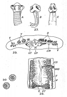
From Alves, etal 2017 (Cit# 7075). Fig. 2 Scanning electron micrographs of representatives of monticelliine proteocephalideans. Chambriella megacephala (Woodland, 1934) ex Sorubimichthys planiceps (MH... MoreFrom Alves, etal 2017 (Cit# 7075). Fig. 2 Scanning electron micrographs of representatives of monticelliine proteocephalideans. Chambriella megacephala (Woodland, 1934) ex Sorubimichthys planiceps (MHNG-PLAT 54608): A, B, Scoleces, dorsoventral and apical views, respectively; C, D, G, Microtriches on the apex of the scolex, external upper surface of the suckers and between suckers, respectively. |
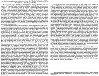
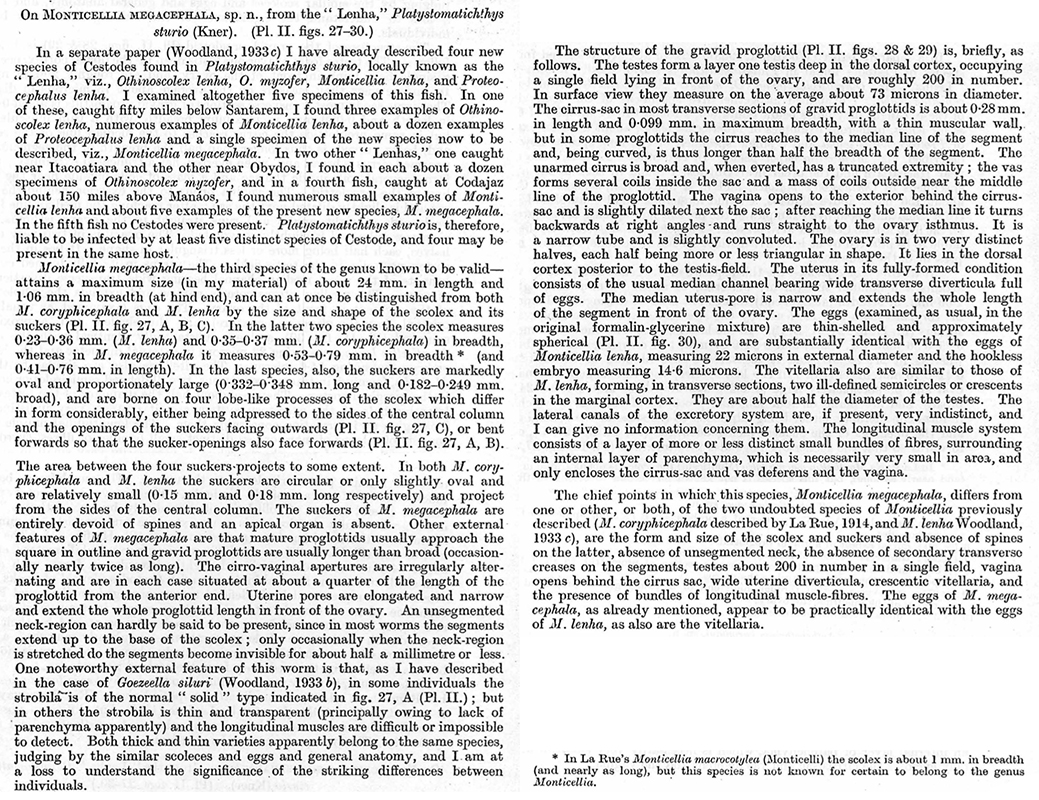
 Pl. II. 27-30. Monticellia megacephala sp. n. Fig. 27. (X17): A, side aspect of scolex, with the four lobes turned forwards (for attachment?), the openings of the four large oval suckers lying inside the circular cavity surrounding the apex and facing forwards; B, side aspect of scolex, with the four lobes turned back, so exposing the four sucker-openings; C, the lobes of this scolex are turned back and in close contact with the base of the scolex. 28 (X27): ventral aspect of gravid proglottid. The uterus is shown as being empty in order not to confuse the drawing. 29 (X56): transverse section of a gravid proglottid through the anterior end of one side of the ovary (oblique section). The subcuticula and parenchyma are not shown. 30 (X395): two undistorted intra-uterine eggs.
 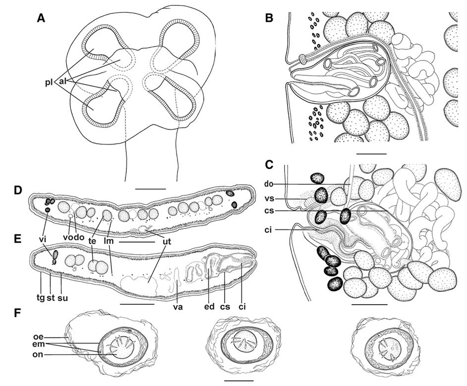 From Alves, etal 2017 (Cit# 7075). Fig. 3 Chambriella megacephala (Woodland, 1934) ex Zungaro jahu. A, Scolex, apical view (paratype of C. agostinhoi, CHIOC 32820c); B, Terminal genitalia, ventral view (holotype of C. agostinhoi, CHIOC 32820a); C, Terminal genitalia, dorsal view (MHNH- PLAT 22025); D, E, Cross-sections at anterior and cirrus-sac level of proglottides, respectively (MHNH-PLAT 19546); F, Eggs drawn in distilled water (MHNH-PLAT 20768). Abbreviations: al, anterior sucker loculus; ci, cirrus; cs, cirrus-sac; do, dorsal osmoregulatory canal; ed, ejaculatory duct; em, bi-layered embryophore; lm, internal longitudinal musculature; oe, outer envelope; on, oncosphere; pl, posterior sucker loculus; st, subtegumental muscle fibres; su, subtegumental cells; te, testes; tg, tegument; ut, uterus; va, vas deferens; vi, vitelline follicles; vo, ventral osmoregulatory canal; vs, vaginal sphincter. Scale-bars: A, D, E, 200 µm; B, C, 100 µm; F, 20 µm
 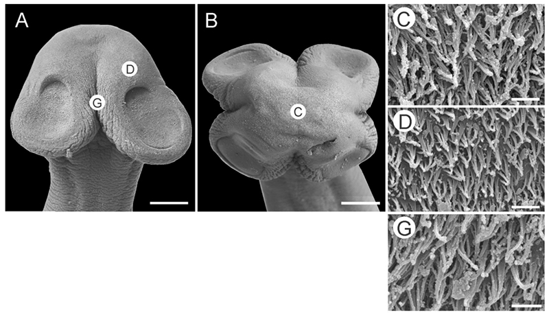 From Alves, etal 2017 (Cit# 7075). Fig. 2 Scanning electron micrographs of representatives of monticelliine proteocephalideans. Chambriella megacephala (Woodland, 1934) ex Sorubimichthys planiceps (MHNG-PLAT 54608): A, B, Scoleces, dorsoventral and apical views, respectively; C, D, G, Microtriches on the apex of the scolex, external upper surface of the suckers and between suckers, respectively.