Cestode Scientific Name
| Species ID | 2536 |
|---|---|
| Order | Onchoproteocephalidea I |
| Family | Proteocephalidae |
| Subfamily | Proteocephalinae |
| Genus | Batrachotaenia |
| Species | saphena |
| Authority | (Osler, 1931) Freze, 1965 |
| Taxonomic Status | Synonym |
| Valid Name | Ophiotaenia saphena Osler, 1931 |
| Synonyms | Crepidobothrium saphena (Osler, 1931) Ingles, 1936; Proteocephalus saphenus (Osler, 1931) Brooks, 1978 |
| Genus Record | No |
| Type Species | No |
| Verified | Yes |
| Verified By | T. Scholz |
| Citation(s) |
Osler, C. P. 1931. A new cestode from Rana clamitans Latr.. Journal of Parasitology 17(4): 183-186. (4147) Download PDFFreze, V. I. 1965. [Proteocephalata in Fish, Amphibians, and Reptiles] (In Russian). Osnovy Tsestodologii 5: 538 pp. (4031) Download PDF |
| Redescription | |
| Scientific Name Notes |
Record Data
| Date (MM/DD/YYYY) | Action | User Name |
|---|---|---|
| 03/21/2003 | Created | Scholz, Kuchta, Healy, Caira |
| 02/09/2010 | Modified | |
| 02/21/2020 | Modified | P. Lopez Dineen |
| 04/15/2020 | Modified | T. Scholz |
| 07/22/2020 | Modified | B. Barbeau |
| 12/03/2021 | Modified | B. Barbeau |
Type Host
| Host Class | |||||||
|---|---|---|---|---|---|---|---|
| Host Order | |||||||
| Host Family | |||||||
|
Type Host (Literal) |
|
||||||
|
Type Host (Valid) |
|
||||||
| Additional Host(s) | |||||||
| Site in Host | |||||||
| Host Notes |
Type Locality
| Country | |
|---|---|
| Body of Water | |
| Island(s) | |
| City/Region | |
| Coordinates | |
| DD Latitude | |
| DD Longitude | |
| Additional Localities | |
| Locality Notes |
Specimens
| Type Material | |
|---|---|
| Total Number of Type Specimens | |
| Voucher Material | |
| Specimen Notes |
Data are given as in original description unless otherwise indicated.

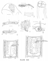
 All drawings were made with the camera lucida. The projected scale in figures 1 and 7 represents 0.02mm; in all other figures, 0.2mm. Fig. 1. - Scolex of adult cestode; longitudinal section showing vestigial fifth sucker with basement membrane, nuclei, muscle fibers, tubular opening. Partial outlines of two other suckers are shown for reference. Fig. 2. - Ventral aspect of protruded cirrus and vagina, showing ductus ejaculatorius coiled. Fig. 3. - First proglottids. Fig. 4. - Scolex of adult cestode from a toto moutn. Fig. 5. - Cross-section through gravid proglottid at level of cirrus-pouch, showing longitudinal muscles, vitellaria, lateral nerve, ventral excretory tube, testes, uterus, vas deferens, cirrus pouch and cirrus. Fig. 6. - Cross-section through same proglottid as in Fig. 5, at level of ovary. Fig. 7. - Oötype with Mehlis' gland (shell gland), oviduct, and beginning of uterine passage; frontal section. Fig 8. - Organs of interovarial space reconstructed from camera outlines. Dorsal side uppermost. Fig. 9. - Mature proglottid, ventral aspect. Fig. 10. - Ruptured proglottid, ventral aspect. Abbreviations Used. c. Cirrus. e. Ventral excretory tube. i. Interovarian organs. o. Ovary. u. Uterus. v. Vitelline duct.

Best viewed in Firefox


