Line Drawing 1
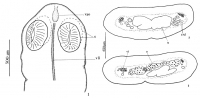
FIGURES 1-5. Drawings of Proteocephalus chamelensis n. sp. 1. Scolex. 2, 3. Cross sections of gravid proglottides showing the medullary position of vitelline follicles and testes. 4. Mature proglottis... MoreFIGURES 1-5. Drawings of Proteocephalus chamelensis n. sp. 1. Scolex. 2, 3. Cross sections of gravid proglottides showing the medullary position of vitelline follicles and testes. 4. Mature proglottis. 5. Gravid proglottis; cp, cirrus pouch; exd, excretory duct; eyd, ejaculatory duct; o, ovary; pu, preformed uterus; s, suckers; t, testes; u, uterus; v, vagina; vao, vestigial apical organ; vd, vas deferens; vi, vitellaria; vli, ventral longitudinal incision |
Line Drawing 2

FIGURES 4-5. Drawings of Proteocephalus chamelensis n. sp. 4. Mature proglottis. 5. Gravid proglottis; cp, cirrus pouch; exd, excretory duct; eyd, ejaculatory duct; o, ovary; pu, preformed uterus; s, ... MoreFIGURES 4-5. Drawings of Proteocephalus chamelensis n. sp. 4. Mature proglottis. 5. Gravid proglottis; cp, cirrus pouch; exd, excretory duct; eyd, ejaculatory duct; o, ovary; pu, preformed uterus; s, suckers; t, testes; u, uterus; v, vagina; vao, vestigial apical organ; vd, vas deferens; vi, vitellaria; vli, ventral longitudinal incision |
Photo Micrograph
|
Scanning Electron Micrograph
|


 FIGURES 1-5. Drawings of Proteocephalus chamelensis n. sp. 1. Scolex. 2, 3. Cross sections of gravid proglottides showing the medullary position of vitelline follicles and testes. 4. Mature proglottis. 5. Gravid proglottis; cp, cirrus pouch; exd, excretory duct; eyd, ejaculatory duct; o, ovary; pu, preformed uterus; s, suckers; t, testes; u, uterus; v, vagina; vao, vestigial apical organ; vd, vas deferens; vi, vitellaria; vli, ventral longitudinal incision
 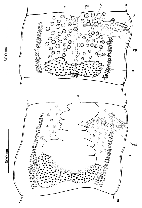 FIGURES 4-5. Drawings of Proteocephalus chamelensis n. sp. 4. Mature proglottis. 5. Gravid proglottis; cp, cirrus pouch; exd, excretory duct; eyd, ejaculatory duct; o, ovary; pu, preformed uterus; s, suckers; t, testes; u, uterus; v, vagina; vao, vestigial apical organ; vd, vas deferens; vi, vitellaria; vli, ventral longitudinal incision


