Line Drawing 1
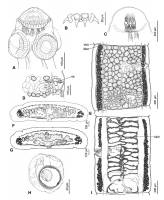
Figure 4. Gangesia agraensis Verma, 1928 from Wallago attu, India. A. Scolex, dorsoventral view (IPCAS C-617, field no. IND 167). B. Rostellar hooks (MHNG-PLAT 82298, field no. IND 795B). C. Detail of... MoreFigure 4. Gangesia agraensis Verma, 1928 from Wallago attu, India. A. Scolex, dorsoventral view (IPCAS C-617, field no. IND 167). B. Rostellar hooks (MHNG-PLAT 82298, field no. IND 795B). C. Detail of retractor muscles. D. Terminal genitalia, dorsal view (IPCAS C-617, field no. IND 795). E. Mature proglottis, dorsal view (IPCAS C-617, field no. IND 167). F, G. Cross sections at level of testicular field and ovary, respectively (MHNG-PLAT 60725, field no. 117/08). H. Egg drawn in distilled water. I. Gravid proglottis, ventral view (IPCAS C-617, field no. IND 795). Abbreviations: doc - dorsal osmoregulatory canal, upo - uterine pore- like opening, voc - ventral osmoregulatory canal, vs - vaginal sphincter. |
Line Drawing 2
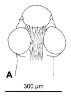
Figure 9. Outline of scoleces with retractor muscles of selected Gangesia taxa. A. Gangesia agraensis. |
Photo Micrograph
|
Scanning Electron Micrograph
Figure 5. Scanning electron photomicrographs of the scolex of Gangesia agraensis Verma, 1928. A. Scolex, dorsoventral view. B. Scolex apical view. C. Suckers with small hooklets on the outer rim. D. D... MoreFigure 5. Scanning electron photomicrographs of the scolex of Gangesia agraensis Verma, 1928. A. Scolex, dorsoventral view. B. Scolex apical view. C. Suckers with small hooklets on the outer rim. D. Detail of rostellum-like organ, dorsoventral view; note on row of rostellar hooks. E. Detail of the outer rim of the sucker with hooklets. F. Detail of papilliform filitriches on the rostellum-like organ. G. Detail of papilliform filitriches on the sucker. H. Detail of acicular filitriches in between the suckers. I. Detail of capilliform filitriches and small gladiate spinitriches on the neck region. Scale bars: A -100 µm; B-D -20 µm; E, I -2 µm; F-G -1µm. |


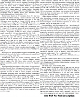
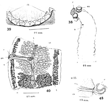
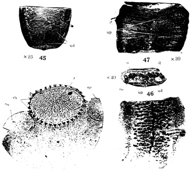
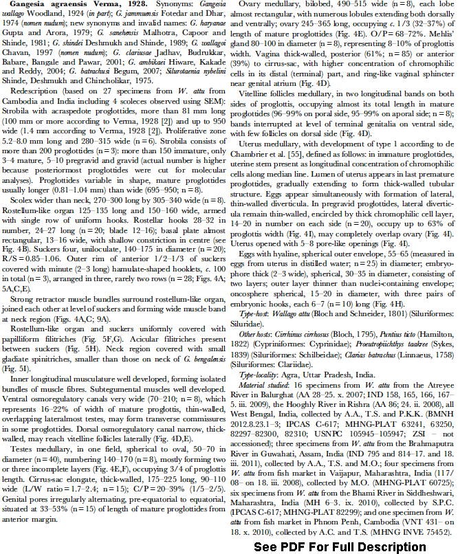
 Figure 4. Gangesia agraensis Verma, 1928 from Wallago attu, India. A. Scolex, dorsoventral view (IPCAS C-617, field no. IND 167). B. Rostellar hooks (MHNG-PLAT 82298, field no. IND 795B). C. Detail of retractor muscles. D. Terminal genitalia, dorsal view (IPCAS C-617, field no. IND 795). E. Mature proglottis, dorsal view (IPCAS C-617, field no. IND 167). F, G. Cross sections at level of testicular field and ovary, respectively (MHNG-PLAT 60725, field no. 117/08). H. Egg drawn in distilled water. I. Gravid proglottis, ventral view (IPCAS C-617, field no. IND 795). Abbreviations: doc - dorsal osmoregulatory canal, upo - uterine pore- like opening, voc - ventral osmoregulatory canal, vs - vaginal sphincter.
  Figure 9. Outline of scoleces with retractor muscles of selected Gangesia taxa. A. Gangesia agraensis.
  Figure 5. Scanning electron photomicrographs of the scolex of Gangesia agraensis Verma, 1928. A. Scolex, dorsoventral view. B. Scolex apical view. C. Suckers with small hooklets on the outer rim. D. Detail of rostellum-like organ, dorsoventral view; note on row of rostellar hooks. E. Detail of the outer rim of the sucker with hooklets. F. Detail of papilliform filitriches on the rostellum-like organ. G. Detail of papilliform filitriches on the sucker. H. Detail of acicular filitriches in between the suckers. I. Detail of capilliform filitriches and small gladiate spinitriches on the neck region. Scale bars: A -100 µm; B-D -20 µm; E, I -2 µm; F-G -1µm.

