Line Drawing 1
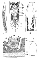
FIGURES 1 TO 5. Host: Catostomus commersoni (Lacepede) unless otherwise specified. Locality: New York, USA, unless otherwise specified. Abbreviations: AEC, ascending excretory canal; CML, circular mus... MoreFIGURES 1 TO 5. Host: Catostomus commersoni (Lacepede) unless otherwise specified. Locality: New York, USA, unless otherwise specified. Abbreviations: AEC, ascending excretory canal; CML, circular muscle layer; DEC, descending excre- tory canal; E, epithelium; EB, excretory bladder; ESV, extemal seminal vesicle; FGp, female gonopore; GpOt, glandular portion ootype; ILM, inner longitudinal muscles; L, layer; LG, longitudinal groove; LML, longitudinal muscle layer; MGp, male gonopore; 0, ovary; OLM, outer longitudinal muscles; Op, operculum; Ov, ovum; SE, subepithelium; T, testis; TI, terminal introvert; U, uterus; V, vitellarium; Va, vagina; VD, vas deferens. Figure 1. Mature worm, toto, holotype. Figure 2. Reproductive systems, toto, holotype. Figure 3. Scolex, holotype. Figure 4. Scolex, in situ, sagittal section. Figure 5. Extended scolex of immature specimen. |
Line Drawing 2

FIGURES 6 TO 13. Figure 6. Scolex, M. ingens Hunter, from Ictiobus cyprinellus, St. Croix River, Minnesota. Collection of T. Jensen. Figure 7. Scolex, M. wageneri Nybelin from Tinca tinca (L.), River ... MoreFIGURES 6 TO 13. Figure 6. Scolex, M. ingens Hunter, from Ictiobus cyprinellus, St. Croix River, Minnesota. Collection of T. Jensen. Figure 7. Scolex, M. wageneri Nybelin from Tinca tinca (L.), River Odra, Poland. Collection of J. Janiszewska. Figure 8. Reproductive systems, sagittal section. Figure 9. Body, cross section. Figure 10. Scolex, showing maximum neck constriction. Figure 11. Whole worms, variations in shape of fixed specimens. Figure 12. Egg. Figure 13. Immature worm, toto. |
Photo Micrograph
|
Scanning Electron Micrograph
|
 FIGURES 1 TO 5. Host: Catostomus commersoni (Lacepede) unless otherwise specified. Locality: New York, USA, unless otherwise specified. Abbreviations: AEC, ascending excretory canal; CML, circular muscle layer; DEC, descending excre- tory canal; E, epithelium; EB, excretory bladder; ESV, extemal seminal vesicle; FGp, female gonopore; GpOt, glandular portion ootype; ILM, inner longitudinal muscles; L, layer; LG, longitudinal groove; LML, longitudinal muscle layer; MGp, male gonopore; 0, ovary; OLM, outer longitudinal muscles; Op, operculum; Ov, ovum; SE, subepithelium; T, testis; TI, terminal introvert; U, uterus; V, vitellarium; Va, vagina; VD, vas deferens. Figure 1. Mature worm, toto, holotype. Figure 2. Reproductive systems, toto, holotype. Figure 3. Scolex, holotype. Figure 4. Scolex, in situ, sagittal section. Figure 5. Extended scolex of immature specimen.
 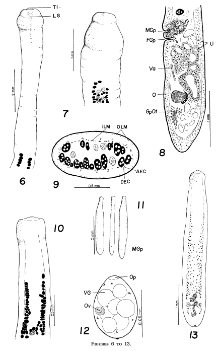 FIGURES 6 TO 13. Figure 6. Scolex, M. ingens Hunter, from Ictiobus cyprinellus, St. Croix River, Minnesota. Collection of T. Jensen. Figure 7. Scolex, M. wageneri Nybelin from Tinca tinca (L.), River Odra, Poland. Collection of J. Janiszewska. Figure 8. Reproductive systems, sagittal section. Figure 9. Body, cross section. Figure 10. Scolex, showing maximum neck constriction. Figure 11. Whole worms, variations in shape of fixed specimens. Figure 12. Egg. Figure 13. Immature worm, toto.

