Line Drawing 1

Figs. 1-5. Andrepigynotaenia haematopodis, n.g., n.sp. Fig. 1. Scolex. Fig. 2. Complete strobila, showing the female genitalia developing before the male. Fig. 3. Cirrus-sac and invaginated cirrus. Fi... MoreFigs. 1-5. Andrepigynotaenia haematopodis, n.g., n.sp. Fig. 1. Scolex. Fig. 2. Complete strobila, showing the female genitalia developing before the male. Fig. 3. Cirrus-sac and invaginated cirrus. Fig. 4. Cirrus-sac and evaginated cirrus. Fig. 5. One of the hooks of the cirrus. Figs. 6-8. Andrepigynotaenia haematopodis, n.g., n.sp. Fig. 6. Two adjacent proglottides, showing functional female genitalia. Dorsal view. Fig. 7. Proglottis, showing functional male organs, gravid uterus and remains of receptaculum seminis. Dorsal view. Fig. 8. Gravid proglottis containing uterus, cirrus-sac, degenerating testes and receptaculum seminis. Dorsal view. |
Line Drawing 2
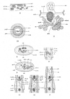
Figs. 9-11. Andrepigynotaenia haematopodis, n.g., n.sp. Fig. 9. Diagram of portion of musculature in transverse section. Fig. 10. Transverse section through cirrus-sac. Fig. 11. Reconstruction of fema... MoreFigs. 9-11. Andrepigynotaenia haematopodis, n.g., n.sp. Fig. 9. Diagram of portion of musculature in transverse section. Fig. 10. Transverse section through cirrus-sac. Fig. 11. Reconstruction of female genitalia; vitellarium displaced slightly to the right. Dorsal view. Figs. 12-17. AndJrepigynotaenia haematopodis, n.g.,n.sp. Fig. 12. Transverse section through proglottis, showing U-shaped ovary. Fig. 13. Transverse section through proglottis, showing arrangement of some of female organs and
ducts, and developing cirrus passing dorsally to both lateral excretory vessels. Fig. 14, Sagittal section, showing cirrus-sac dorsal to excretory vessels. Fig. 15. Sagittal section, showing junction of sperm-duct with oviduct, etc. Fig. 16. Sagittal section, showing vitellarium, vitelline duct, etc. Fig. 17. Sagittal section, showing course of uterus from ootype to segment next in front. |
Photo Micrograph
|
Scanning Electron Micrograph
|

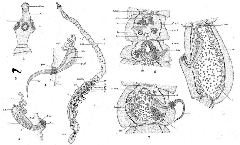 Figs. 1-5. Andrepigynotaenia haematopodis, n.g., n.sp. Fig. 1. Scolex. Fig. 2. Complete strobila, showing the female genitalia developing before the male. Fig. 3. Cirrus-sac and invaginated cirrus. Fig. 4. Cirrus-sac and evaginated cirrus. Fig. 5. One of the hooks of the cirrus. Figs. 6-8. Andrepigynotaenia haematopodis, n.g., n.sp. Fig. 6. Two adjacent proglottides, showing functional female genitalia. Dorsal view. Fig. 7. Proglottis, showing functional male organs, gravid uterus and remains of receptaculum seminis. Dorsal view. Fig. 8. Gravid proglottis containing uterus, cirrus-sac, degenerating testes and receptaculum seminis. Dorsal view.
 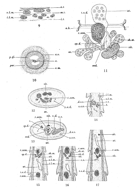 Figs. 9-11. Andrepigynotaenia haematopodis, n.g., n.sp. Fig. 9. Diagram of portion of musculature in transverse section. Fig. 10. Transverse section through cirrus-sac. Fig. 11. Reconstruction of female genitalia; vitellarium displaced slightly to the right. Dorsal view. Figs. 12-17. AndJrepigynotaenia haematopodis, n.g.,n.sp. Fig. 12. Transverse section through proglottis, showing U-shaped ovary. Fig. 13. Transverse section through proglottis, showing arrangement of some of female organs and
ducts, and developing cirrus passing dorsally to both lateral excretory vessels. Fig. 14, Sagittal section, showing cirrus-sac dorsal to excretory vessels. Fig. 15. Sagittal section, showing junction of sperm-duct with oviduct, etc. Fig. 16. Sagittal section, showing vitellarium, vitelline duct, etc. Fig. 17. Sagittal section, showing course of uterus from ootype to segment next in front.
Figs. 9-11. Andrepigynotaenia haematopodis, n.g., n.sp. Fig. 9. Diagram of portion of musculature in transverse section. Fig. 10. Transverse section through cirrus-sac. Fig. 11. Reconstruction of female genitalia; vitellarium displaced slightly to the right. Dorsal view. Figs. 12-17. AndJrepigynotaenia haematopodis, n.g.,n.sp. Fig. 12. Transverse section through proglottis, showing U-shaped ovary. Fig. 13. Transverse section through proglottis, showing arrangement of some of female organs and
ducts, and developing cirrus passing dorsally to both lateral excretory vessels. Fig. 14, Sagittal section, showing cirrus-sac dorsal to excretory vessels. Fig. 15. Sagittal section, showing junction of sperm-duct with oviduct, etc. Fig. 16. Sagittal section, showing vitellarium, vitelline duct, etc. Fig. 17. Sagittal section, showing course of uterus from ootype to segment next in front.