Cestode Scientific Name
| Species ID | 192 |
|---|---|
| Order | Caryophyllidea |
| Family | Caryophyllaeidae |
| Subfamily | |
| Genus | Khawia |
| Species | baltica |
| Authority | Szidat, 1942 |
| Taxonomic Status | Synonym |
| Valid Name | Caryophyllaeus balticus (Szidat, 1942) Barčák, Scholz, Oros & Hanzelová, 2017 |
| Synonyms | |
| Genus Record | No |
| Type Species | No |
| Verified | Yes |
| Verified By | T. Scholz |
| Citation(s) |
Szidat, L.. 1942. Über die Caryophyllaeiden-Gattung Khawia H. F. Hsü, 1935 und eine neue Art dieser Gattung, Khawia baltica n. spec. . Zeitschrift für Parasitenkunde 12: 120-132. (701) Download PDFBarčák, D., M. Oros, V. Hanzelová, and T. Scholz. 2017. A synoptic review of Caryophyllaeus Gmelin, 1790 (Cestoda: Caryophyllidea), parasites of cyprinid fishes. Folia Parasitologica 64: 027. (7142) Download PDF |
| Redescription |
Scholz, T., J. Brabec, I. Král'ová-Hromadová, M. Oros, E. Bazsalovicsová, A. Ermolenko, V. Hanzelová. 2011. Revision of Khawia spp. (Cestoda: Caryophyllidea), parasites of cyprinid fish, including a key to their identification and molecular phylogeny. Folia Parasitologica 58(3): 197-223. (6186) Download PDF |
| Scientific Name Notes | The year of species description should be 1942 because the description appeared in this year, not in 1941 as generally though, including Schmidt (1986) |
Record Data
| Date (MM/DD/YYYY) | Action | User Name |
|---|---|---|
| 10/14/2005 | Created | Scholz, Oros |
| 02/04/2010 | Modified | |
| 11/18/2015 | Modified | N. Arisco |
| 02/08/2016 | Modified | J. Caira |
| 05/04/2016 | Modified | B. Barbeau |
| 08/07/2017 | Modified | M. Oros |
| 04/03/2020 | Modified | T. Scholz |
Type Host
| Host Class | |||||||
|---|---|---|---|---|---|---|---|
| Host Order | |||||||
| Host Family | |||||||
|
Type Host (Literal) |
|
||||||
|
Type Host (Valid) |
|
||||||
| Additional Host(s) | |||||||
| Site in Host | small intestine | ||||||
| Host Notes |
Type Locality
| Country | East Prussia [Russia] |
|---|---|
| Body of Water | |
| Island(s) | |
| City/Region | Rossitten, Kurischen Nehrung |
| Coordinates | |
| DD Latitude | |
| DD Longitude | |
| Additional Localities | |
| Locality Notes |
Specimens
| Type Material | |
|---|---|
| Total Number of Type Specimens | |
| Voucher Material | |
| Specimen Notes | Deposition of type material not indicated |
Data are given as in original description unless otherwise indicated.
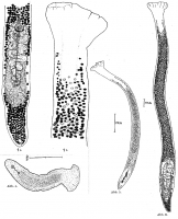
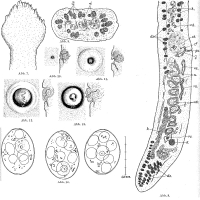
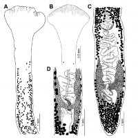
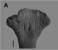

 Abb. 1a und b. Khawia baltica n. spec. a Letztes Körperviertel mit dem Geschlechtsapparat. b Vorderende mit Scolex. Abb. 4. Khawia baltica n. spec. Jüngstes Stadium aus dem Darm von Tinca tinca. Abb. 5. Khawia baltica n. spec. Junger Wurm mit protandrisch reifenden Geschlechtsdrüsen. Abb. 6. Khawia baltica n. spec. Totalansicht des ausgewachsenen Wurmes.
  Abb. 7. Khawia baltica n. spec. Scolex in Bewegung. Nach dem Leben gezeichnet. Abb. 8. Khawia baltica n. spec. Sagittalschnitt durch das Hinterende. cb = Cirrusbentel, do - Dotterstocksfollikel, ga = Genitalatrium, h = Hodenbläschen, k = Ovarium, rs = Receptaculum seminis, u = Uterusschlingen, udr = Uterusdrüsen, va = Vagina, vd = Vas deferens. Abb. 9. Khawia baltica n. spec. Querschnitt durch den mittleren Teil des Körpers mit Hodenbläschen und Dotterstocksfollikeln. Abb. 10-13. Khawia baltica n. spec. Bildung der männlichen Genitalpapille in vier verschiedenen Phasen in Aufsicht und Sagittalschnitt. Nach dem Leben. Abb. 14. Khawia baltica n. spec. Eier aus dem Darminhalt von Tinca tinca.
 Fig. 3. Khawia baltica Szidat, 1941 from Tinca tinca, Czech Republic (IPCAS C-42). A, B - anterior part of the body (note the presence of grooves on the anterior margin of the scolex and position of the first vitelline follicles and testes - in A, the latter situated very far posterior to vitelline follicles); C - posterior part of the body, dorsal (note the presence of numerous vitelline follicles alongside the preovarian uterine loops); D - ovarian region, ventral (note variation in the number of vitelline follicles alongside the ovarian arms).
  Fig. 8. Khawia baltica Szidat, 1941 from Tinca tinca, Czech Republic (A) Scanning electron micrographs of scoleces. Scale bars: 200 µm (A) and 100 µm (B).
 Best viewed in Firefox


