Cestode Scientific Name
| Species ID | 180 |
|---|---|
| Order | Caryophyllidea |
| Family | Capingentidae |
| Subfamily | |
| Genus | Glaridacris |
| Species | terebrans |
| Authority | (Linton, 1893) Mackiewicz, 1974 |
| Taxonomic Status | Valid |
| Valid Name | |
| Synonyms | Monobothrium terebrans Linton, 1893 Caryophyllaeus terebrans (Linton, 1893) Woodland, 1923 |
| Genus Record | No |
| Type Species | No |
| Verified | Yes |
| Verified By | T. Scholz |
| Citation(s) |
Linton, E. 1893. On fish entozoa from Yellowstone national park. Report of the U.S. Commissioner of Fish and Fisheries for 1889-1891 pt. 17: 545-564. (6409) Download PDFMackiewicz, J. S. 1974. The genus Caryophyllaeus Gmelin (Cestoidea: Caryophyllidea) in the Nearctic. Proceedings of the Helminthological Society of Washington 41: 184-191. (695) Download PDF |
| Redescription |
Oros, M., D. Uhrovič, and T. Scholz . 2018. A new classification of Glaridacris Cooper, 1920 (Cestoda: Caryophyllidea), parasites of suckers (Catostomidae) in north America, including erection of Pseudoglaridacris n.gen.. Journal of Parasitology 104: 60-69.. (7166) Download PDF |
| Scientific Name Notes |
Record Data
| Date (MM/DD/YYYY) | Action | User Name |
|---|---|---|
| 10/14/2005 | Created | Scholz, Oros, V. Lopez |
| 07/25/2014 | Modified | |
| 05/11/2016 | Modified | B. Barbeau |
| 04/02/2020 | Modified | T. Scholz |
| 08/12/2021 | Modified | T. Scholz |
Type Host
| Host Class | Actinopterygii | ||||||
|---|---|---|---|---|---|---|---|
| Host Order | Cypriniformes | ||||||
| Host Family | Catostomidae | ||||||
|
Type Host (Literal) |
|
||||||
|
Type Host (Valid) |
|
||||||
| Additional Host(s) | |||||||
| Site in Host | intestine | ||||||
| Host Notes |
Type Locality
| Country | U.S.A. |
|---|---|
| Body of Water | Heart Lake |
| Island(s) | |
| City/Region | Wyoming |
| Coordinates | |
| DD Latitude | |
| DD Longitude | |
| Additional Localities | |
| Locality Notes |
Specimens
| Type Material | |
|---|---|
| Total Number of Type Specimens | |
| Voucher Material | |
| Specimen Notes |
Data are given as in original description unless otherwise indicated.


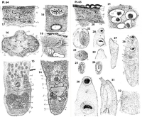
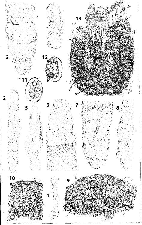 Plate 63. Monobothrium terebrans sp. nov. Fig. 1. Adult; a, head; b, posterior end; c, genital pore, X 34. Fig. 2. Smaller specimen, dorsal view, X 18. Fig. 3. Posterior end of adult, ventral view; a, genital pore, X 14. Fig. 4. Small specimen, dorsal view, X 14. Fig. 5. Anterior end of adult, marginal view, X 14. Fig. 6. Anterior end of adult, dorsal view, X 14. Fig. 7. Posterior end of adult, dorsal view, X 14. Fig. 8. Posterior end of adult, marginal view, X 14. (Figs. 5 to 8 are sketched from the same specimen) Fig. 9. Median longitudinal section of anterior end of small specimen, parallel with a dorsal surface, X 200. Fig. 10. Transverse section near apex, of small specimen; c, cuticle; v, vessel of water-vascular syste, X 200. Fig. 11. Egg, sketched from section of young specimen, showing eggs in the uterus, X 375. Fig. 12. Egg, from one of the posterior convolutions of the uterus of a young specimen; a, germ cell; the remainder of the contents consists of globular masses from the vitelline gland, X 375. Fig. 13. Longitudinal section through the posterior region of a small specimen; c, cuticle; ci, cirrus and cirrus-pouch; e, epidermis; gg, marginal lobes of germ gland; lm, longitudinal muscles; oo, eggs in posterior convolutions of the uterus; sc, subcuticular fibro-granular layer; t, testes; uu, uterus; v, seminal receptacle of vagina; vd, vas deferens; vg vg vg, marginal and posterior vitelline glands, X 300.
 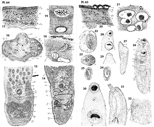 Plate 64. Monobothrium terebrans sp. nov. Fig. 14. Longitudinal section through the posterior region of an adult specimen, X 60; u, shell gland; other letters as in Fig. 13.; Fig. 15. Diagrammatic sketch showing position of genitalia; v, vagina; v', seminal receptacle; g, shell gland; other letters as in Fig. 13.; Fig. 16. Transverse section through body in region of cirrus bulb of adult, X 54; l, longitudinal muscles in subcutaneous fibro-granular layer; w, vessels of water-vascular system; other letters as in Fig. 13.; Fig. 17. Transverse section of body-wall in front of cirrus, X 210; l, longitudinal subcutatneous fibers; other letters as in Fig. 13.; Fig. 18. From transverse section in front of germ gland, X 210; c, cilliated duct vagina; g, germ gland; lm, longitudinal muscles; o, egg in uterus; u, uterus. Fig. 19. Longitudinal section near ventral surface; ci, cirrus; v, vagina, X 300. Plate 65. Monobothrium terebrans sp. nov. Fig. 20. Longitudinal section of body wall near posterior end; e, epidermis; c, cuticle; l, longitudinal subcuticular fibers; sc, subcuticular fibro-granular layers; n, nucleated cell; lm, longitudinal muscle layer; g, nucleated cells of germ gland. X 300. Fig. 21. Longitudinal section through vas deferens; vd, vas deferens; s, masses of spermatozoa, X 375.
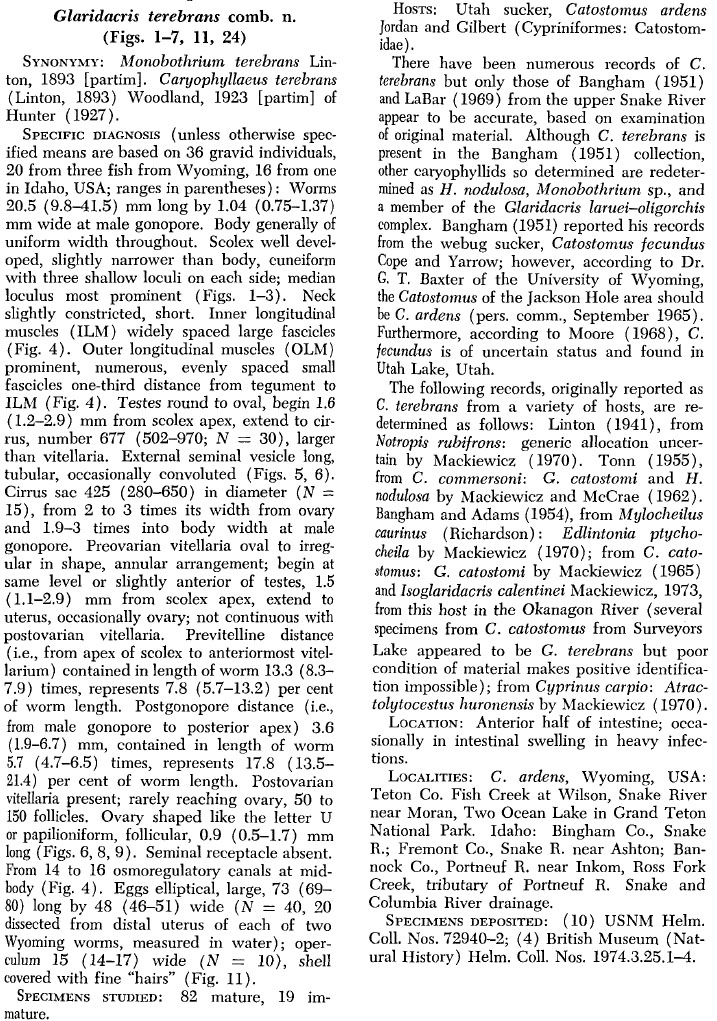
Best viewed in Firefox


