Line Drawing 1
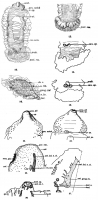
PLATE 34.
Fig. 10. Gyrocotyle fimbriata, ventral. Flattened, stained in borax
carmine, cleared in cedar-oil. Showing arrangement of spines at posterior extremity. Spines at anterior extremity not sh... MorePLATE 34.
Fig. 10. Gyrocotyle fimbriata, ventral. Flattened, stained in borax
carmine, cleared in cedar-oil. Showing arrangement of spines at posterior extremity. Spines at anterior extremity not shown. X 4.
Fig. 12. G. fimbriata, dorsal. Stained flattened specimen. X 4.
Figs. 14, 15. G. fimbriata. Showing canal-opening, with proboscis
inverted, fig. 14, and everted, fig. 15. Sketch without camera.
Fig. 16. Sagittal section of longitudinal nerve-stem, showing sheath cells, branch-nerves, and ganglion-cells of the first order. Iron haematoxylin-erythrosin. X 100.
acet.acetabulum.
can. op.canal opening.
gang. 1st.ganglion cell of the first order.
gen. notchgenital notch.
n.b.nerve branch.
n. fib.nerve fibre.
p. op-penis opening.
par.parenchyma.
post. ros.posterior rosette.
prob.proboscis.
rec. sem.receptaculum seminis.
sh. c.sheath cell.
ut.uterus.
PLATE 36.
Figs. 22, 23. Gyrocotyle fimbriata, anterior extremity, ventral and
dorsal. Showing sensory pits and arrangement of spines. From pre
served specimen. X 13.
Fig. 25. G. fimbriata. Dorsal half of sagittal section of tip of aceta
bulum, showing sensory pit. Borax-carmine, Lyon's blue. X 430.
Fig. 26. G. fimbriata. Sketch, life, acetabular extremity. Showing
anterior excretory ring and deeper-lying ciliated canals.
Fig. 27. G. fimbriata. Sagittal section just lateral of the acetabulum.
Showing anterior longitudinal nerve stem, with branches and anterior ganglionic knot. No histological detail; position of giant-cells within
ganglion knot marked by small circles.
acet. op.acetabular opening.
ant. er. r.anterior excretory ring.
ant. gang. kn.anterior ganglion knot.
ant. lat. m. st.anterior lateral nerve stem.
cil.cilia.
cut.cuticula.
er can.excretory canal.
gen. notch-genital notch.
lat. f.lateral fold.
par. nuc.parenchyma nucleus.
p. op.penis opening.
post. ros.posterior rosette.
sens. pit-sensory pit. -
sp.spine.
test.testis.
ut. po.uterine pore.
test. n.testicular nerve. |
Line Drawing 2
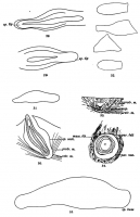
PLATE 37.
Figs. 28, 29. Gyrocotyle fimbriata. Spines, from acetabular group.
Teased out. X 1000.
Fig. 30. G. fimbriata. Spine, from neck of rosette. From specimen
stained in borax-carmine and clea... MorePLATE 37.
Figs. 28, 29. Gyrocotyle fimbriata. Spines, from acetabular group.
Teased out. X 1000.
Fig. 30. G. fimbriata. Spine, from neck of rosette. From specimen
stained in borax-carmine and cleared in cedar-oil. Showing direction of spine and attachment of muscles. X 430.
Fig. 31. G. fimbriata. Spine from neck of rosette. From same speci
men as fig. 30. Drawn in situ. X 430.
Fig. 32. Spines from margin of anterior end, in front of genital notch.
Teased out. X 1000.
Fig. 33. G. fimbriata, transverse section. Cuticula absent. Showing
muscles of spine. X 430. Iron haematoxylin.
Fig. 34. Same as fig. 33. Showing structure of socket of spine.
X 1000.
Fig. 35. Spine from neck of rosette. X 1000.
musc. fib.muscle fibre.
par. feltparenchyma felt.
par. nuc.parenchyma nucleus.
protr. m.protractor muscle.
retr. m.retractor muscle.
sp.spine.
sp. basespine base.
sp. sock.spine socket.
sp. tip-tip of spine. |
Photo Micrograph
|
Scanning Electron Micrograph
|

 PLATE 34.
Fig. 10. Gyrocotyle fimbriata, ventral. Flattened, stained in borax
carmine, cleared in cedar-oil. Showing arrangement of spines at posterior extremity. Spines at anterior extremity not shown. X 4.
Fig. 12. G. fimbriata, dorsal. Stained flattened specimen. X 4.
Figs. 14, 15. G. fimbriata. Showing canal-opening, with proboscis
inverted, fig. 14, and everted, fig. 15. Sketch without camera.
Fig. 16. Sagittal section of longitudinal nerve-stem, showing sheath cells, branch-nerves, and ganglion-cells of the first order. Iron haematoxylin-erythrosin. X 100.
acet.acetabulum.
can. op.canal opening.
gang. 1st.ganglion cell of the first order.
gen. notchgenital notch.
n.b.nerve branch.
n. fib.nerve fibre.
p. op-penis opening.
par.parenchyma.
post. ros.posterior rosette.
prob.proboscis.
rec. sem.receptaculum seminis.
sh. c.sheath cell.
ut.uterus.
PLATE 36.
Figs. 22, 23. Gyrocotyle fimbriata, anterior extremity, ventral and
dorsal. Showing sensory pits and arrangement of spines. From pre
served specimen. X 13.
Fig. 25. G. fimbriata. Dorsal half of sagittal section of tip of aceta
bulum, showing sensory pit. Borax-carmine, Lyon's blue. X 430.
Fig. 26. G. fimbriata. Sketch, life, acetabular extremity. Showing
anterior excretory ring and deeper-lying ciliated canals.
Fig. 27. G. fimbriata. Sagittal section just lateral of the acetabulum.
Showing anterior longitudinal nerve stem, with branches and anterior ganglionic knot. No histological detail; position of giant-cells within
ganglion knot marked by small circles.
acet. op.acetabular opening.
ant. er. r.anterior excretory ring.
ant. gang. kn.anterior ganglion knot.
ant. lat. m. st.anterior lateral nerve stem.
cil.cilia.
cut.cuticula.
er can.excretory canal.
gen. notch-genital notch.
lat. f.lateral fold.
par. nuc.parenchyma nucleus.
p. op.penis opening.
post. ros.posterior rosette.
sens. pit-sensory pit. -
sp.spine.
test.testis.
ut. po.uterine pore.
test. n.testicular nerve.
PLATE 34.
Fig. 10. Gyrocotyle fimbriata, ventral. Flattened, stained in borax
carmine, cleared in cedar-oil. Showing arrangement of spines at posterior extremity. Spines at anterior extremity not shown. X 4.
Fig. 12. G. fimbriata, dorsal. Stained flattened specimen. X 4.
Figs. 14, 15. G. fimbriata. Showing canal-opening, with proboscis
inverted, fig. 14, and everted, fig. 15. Sketch without camera.
Fig. 16. Sagittal section of longitudinal nerve-stem, showing sheath cells, branch-nerves, and ganglion-cells of the first order. Iron haematoxylin-erythrosin. X 100.
acet.acetabulum.
can. op.canal opening.
gang. 1st.ganglion cell of the first order.
gen. notchgenital notch.
n.b.nerve branch.
n. fib.nerve fibre.
p. op-penis opening.
par.parenchyma.
post. ros.posterior rosette.
prob.proboscis.
rec. sem.receptaculum seminis.
sh. c.sheath cell.
ut.uterus.
PLATE 36.
Figs. 22, 23. Gyrocotyle fimbriata, anterior extremity, ventral and
dorsal. Showing sensory pits and arrangement of spines. From pre
served specimen. X 13.
Fig. 25. G. fimbriata. Dorsal half of sagittal section of tip of aceta
bulum, showing sensory pit. Borax-carmine, Lyon's blue. X 430.
Fig. 26. G. fimbriata. Sketch, life, acetabular extremity. Showing
anterior excretory ring and deeper-lying ciliated canals.
Fig. 27. G. fimbriata. Sagittal section just lateral of the acetabulum.
Showing anterior longitudinal nerve stem, with branches and anterior ganglionic knot. No histological detail; position of giant-cells within
ganglion knot marked by small circles.
acet. op.acetabular opening.
ant. er. r.anterior excretory ring.
ant. gang. kn.anterior ganglion knot.
ant. lat. m. st.anterior lateral nerve stem.
cil.cilia.
cut.cuticula.
er can.excretory canal.
gen. notch-genital notch.
lat. f.lateral fold.
par. nuc.parenchyma nucleus.
p. op.penis opening.
post. ros.posterior rosette.
sens. pit-sensory pit. -
sp.spine.
test.testis.
ut. po.uterine pore.
test. n.testicular nerve. 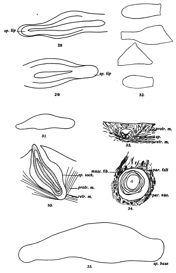 PLATE 37.
Figs. 28, 29. Gyrocotyle fimbriata. Spines, from acetabular group.
Teased out. X 1000.
Fig. 30. G. fimbriata. Spine, from neck of rosette. From specimen
stained in borax-carmine and cleared in cedar-oil. Showing direction of spine and attachment of muscles. X 430.
Fig. 31. G. fimbriata. Spine from neck of rosette. From same speci
men as fig. 30. Drawn in situ. X 430.
Fig. 32. Spines from margin of anterior end, in front of genital notch.
Teased out. X 1000.
Fig. 33. G. fimbriata, transverse section. Cuticula absent. Showing
muscles of spine. X 430. Iron haematoxylin.
Fig. 34. Same as fig. 33. Showing structure of socket of spine.
X 1000.
Fig. 35. Spine from neck of rosette. X 1000.
musc. fib.muscle fibre.
par. feltparenchyma felt.
par. nuc.parenchyma nucleus.
protr. m.protractor muscle.
retr. m.retractor muscle.
sp.spine.
sp. basespine base.
sp. sock.spine socket.
sp. tip-tip of spine.
PLATE 37.
Figs. 28, 29. Gyrocotyle fimbriata. Spines, from acetabular group.
Teased out. X 1000.
Fig. 30. G. fimbriata. Spine, from neck of rosette. From specimen
stained in borax-carmine and cleared in cedar-oil. Showing direction of spine and attachment of muscles. X 430.
Fig. 31. G. fimbriata. Spine from neck of rosette. From same speci
men as fig. 30. Drawn in situ. X 430.
Fig. 32. Spines from margin of anterior end, in front of genital notch.
Teased out. X 1000.
Fig. 33. G. fimbriata, transverse section. Cuticula absent. Showing
muscles of spine. X 430. Iron haematoxylin.
Fig. 34. Same as fig. 33. Showing structure of socket of spine.
X 1000.
Fig. 35. Spine from neck of rosette. X 1000.
musc. fib.muscle fibre.
par. feltparenchyma felt.
par. nuc.parenchyma nucleus.
protr. m.protractor muscle.
retr. m.retractor muscle.
sp.spine.
sp. basespine base.
sp. sock.spine socket.
sp. tip-tip of spine. 

