Line Drawing 1

FIGURES 1-4. Echinobothrium reesae n. sp. 1. Scolex, whowing apical hooks on the rostellum. 2. Group of apical hooks, two small hooks on either side. 3. Scolex, lateral view showing musculature. 4. ... MoreFIGURES 1-4. Echinobothrium reesae n. sp. 1. Scolex, whowing apical hooks on the rostellum. 2. Group of apical hooks, two small hooks on either side. 3. Scolex, lateral view showing musculature. 4. Mature proglottid, ventral view showing reproductive system. |
Line Drawing 2

Fig. 5. The female reprductive complex Key to the Figures. A.H.: Apical hooks, B: Bothridium, Cp: Cephalic peduncle; CS: Cirrus sac, F.C.: Fertilization Canal, G.P.: Genital pore, L.M.: Longitudin... MoreFig. 5. The female reprductive complex Key to the Figures. A.H.: Apical hooks, B: Bothridium, Cp: Cephalic peduncle; CS: Cirrus sac, F.C.: Fertilization Canal, G.P.: Genital pore, L.M.: Longitudinal muscles, MG: Mehlis' gland, OC: Oocapt, OCY: Oocyte, OOT: Ootype, OV: Ovary, OVD: Oviduct, PRM: Protractor muscles, R: Rotellum, RM: Radial muscles, T: Testes, TMR: Transverse muscles of rostellum, UT: Uterus, VAG: Vagina, VD: Vas deferens, VIT: Vitellaria, VIT. D: Vitelline duct |
Photo Micrograph
|
Scanning Electron Micrograph
|

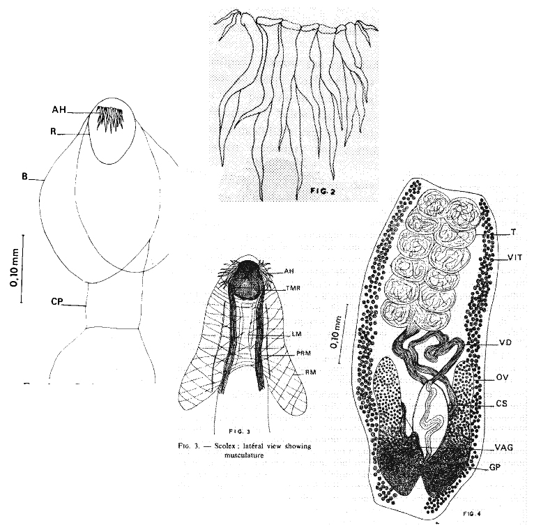 FIGURES 1-4. Echinobothrium reesae n. sp. 1. Scolex, whowing apical hooks on the rostellum. 2. Group of apical hooks, two small hooks on either side. 3. Scolex, lateral view showing musculature. 4. Mature proglottid, ventral view showing reproductive system.
 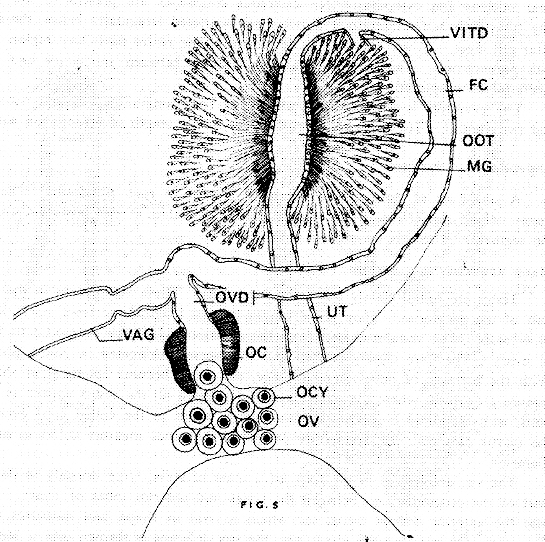 Fig. 5. The female reprductive complex Key to the Figures. A.H.: Apical hooks, B: Bothridium, Cp: Cephalic peduncle; CS: Cirrus sac, F.C.: Fertilization Canal, G.P.: Genital pore, L.M.: Longitudinal muscles, MG: Mehlis' gland, OC: Oocapt, OCY: Oocyte, OOT: Ootype, OV: Ovary, OVD: Oviduct, PRM: Protractor muscles, R: Rotellum, RM: Radial muscles, T: Testes, TMR: Transverse muscles of rostellum, UT: Uterus, VAG: Vagina, VD: Vas deferens, VIT: Vitellaria, VIT. D: Vitelline duct

