Line Drawing 1
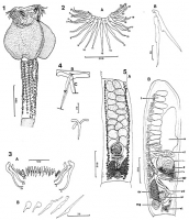
FIGURES 1-5. Echinobothrium raschii n. sp. from Bering Sea skate 1. Scolex, paratype. 2. Rostellar hooks, apical view of large hooks (A) detail of large hooks (B). 3. Rostellar hooklets, continuo... MoreFIGURES 1-5. Echinobothrium raschii n. sp. from Bering Sea skate 1. Scolex, paratype. 2. Rostellar hooks, apical view of large hooks (A) detail of large hooks (B). 3. Rostellar hooklets, continuous row between apical groups of large hooks (A); variation in hooklet form (B). 4. First (anterior) and last (posterior) hooks from row on cephalic peduncle. Interlandmark distances for length (A) and width (B) and posterior extension (C) measurements indicated by brackets on first hook. 5. Proglottid anatomy. (A) Mature proglottid in ventral view; (B) gravid proglottid in lateral view showing ovary in section. (Abreviations: c, cirrus; cs, cirrus sac; gp, gential pore; mg, Mehlis gland; ov, ovary; p, prostate cells; sr, seminal recepticle; t, testis; u, uterus; ud, uterine duct; v, vagina; vd, vas deferens; vt, vitelline follicles. |
Line Drawing 2
|
Photo Micrograph
|
Scanning Electron Micrograph
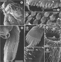
FIGURES 6-13. Scanning electron micrographs of E. raschii. All figures oriented with anterior end toward top of page. 6. Scolex, note large apical hooks of rostellum and microtriches on distal (adhere... MoreFIGURES 6-13. Scanning electron micrographs of E. raschii. All figures oriented with anterior end toward top of page. 6. Scolex, note large apical hooks of rostellum and microtriches on distal (adherent) surface of bothridium. 7. Hooklets forming contiguous row between large apical groups of hooks. 8. Cephalic peduncle. Note that only tips of spines protrude from longitudinal furrows. 9. Detail of rows of palmate microtriches on median distal bothridial surface. 10. Detail of palmate microthrix bearing 15 spinous processes; note small filamentous microtriches covering bothridial surface. 11. Detail of filamentous microtriches and circular pit on proximal bothridial surface. 12. Transition from proximal (right) to distal (left) surfaces of bothridium along posterior bothridial margin; note absence of palmate microtriches on proximal surface (right). 13. Detail of distal bothridial surface on lateral margin showing palmate microtriches; porelike openings are visible where microtriches were attached. Scale bars Figures 5 and 8, 100 µm; scale bars in Figures 7, 9, 12, and 13, 10 µm; scale bar in Figures 10, 11, 1 µm. |
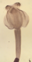
 FIGURES 1-5. Echinobothrium raschii n. sp. from Bering Sea skate 1. Scolex, paratype. 2. Rostellar hooks, apical view of large hooks (A) detail of large hooks (B). 3. Rostellar hooklets, continuous row between apical groups of large hooks (A); variation in hooklet form (B). 4. First (anterior) and last (posterior) hooks from row on cephalic peduncle. Interlandmark distances for length (A) and width (B) and posterior extension (C) measurements indicated by brackets on first hook. 5. Proglottid anatomy. (A) Mature proglottid in ventral view; (B) gravid proglottid in lateral view showing ovary in section. (Abreviations: c, cirrus; cs, cirrus sac; gp, gential pore; mg, Mehlis gland; ov, ovary; p, prostate cells; sr, seminal recepticle; t, testis; u, uterus; ud, uterine duct; v, vagina; vd, vas deferens; vt, vitelline follicles.
 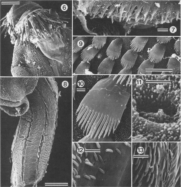 FIGURES 6-13. Scanning electron micrographs of E. raschii. All figures oriented with anterior end toward top of page. 6. Scolex, note large apical hooks of rostellum and microtriches on distal (adherent) surface of bothridium. 7. Hooklets forming contiguous row between large apical groups of hooks. 8. Cephalic peduncle. Note that only tips of spines protrude from longitudinal furrows. 9. Detail of rows of palmate microtriches on median distal bothridial surface. 10. Detail of palmate microthrix bearing 15 spinous processes; note small filamentous microtriches covering bothridial surface. 11. Detail of filamentous microtriches and circular pit on proximal bothridial surface. 12. Transition from proximal (right) to distal (left) surfaces of bothridium along posterior bothridial margin; note absence of palmate microtriches on proximal surface (right). 13. Detail of distal bothridial surface on lateral margin showing palmate microtriches; porelike openings are visible where microtriches were attached. Scale bars Figures 5 and 8, 100 µm; scale bars in Figures 7, 9, 12, and 13, 10 µm; scale bar in Figures 10, 11, 1 µm.
  USNPC No. 86770, paratype slide
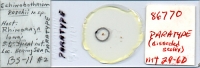


