Cestode Scientific Name
| Species ID | 1695 |
|---|---|
| Order | Diphyllidea |
| Family | |
| Subfamily | |
| Genus | Echinobothrium |
| Species | acanthinophyllum |
| Authority | Rees, 1961 |
| Taxonomic Status | Valid |
| Valid Name | |
| Synonyms | |
| Genus Record | No |
| Type Species | No |
| Verified | Yes |
| Verified By | V.A. Ivanov, R. Kuchta |
| Citation(s) |
Rees, G. 1961. Echinobothrium acanthinophyllum, n. sp. from the spiral valve of Raja montagui Fowler. Parasitology 51: 407-414. (363) Download PDF |
| Redescription | |
| Scientific Name Notes | For more details on this species see Tyler (2006) |
Record Data
| Date (MM/DD/YYYY) | Action | User Name |
|---|---|---|
| 09/09/2006 | Created | K. Jensen , R. Tracy, V. Lopez |
| 08/20/2014 | Modified | |
| 02/19/2016 | Modified | B. Barbeau |
| 08/23/2016 | Modified | R. Kuchta |
Type Host
| Host Class | Chondrichthyes | ||||||
|---|---|---|---|---|---|---|---|
| Host Order | Rajiformes | ||||||
| Host Family | Rajidae | ||||||
|
Type Host (Literal) |
|
||||||
|
Type Host (Valid) |
|
||||||
| Additional Host(s) | |||||||
| Site in Host | spiral intestine | ||||||
| Host Notes |
Type Locality
| Country | England |
|---|---|
| Body of Water | English Channel |
| Island(s) | |
| City/Region | Plymouth |
| Coordinates | |
| DD Latitude | |
| DD Longitude | |
| Additional Localities | |
| Locality Notes |
Specimens
| Type Material | BMNH No. 1962.28.14 (holotype) |
|---|---|
| Total Number of Type Specimens | 1 specimen |
| Voucher Material | |
| Specimen Notes |
Data are given as in original description unless otherwise indicated.
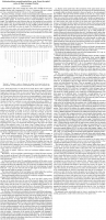
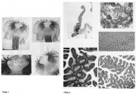
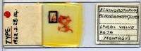
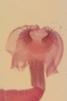
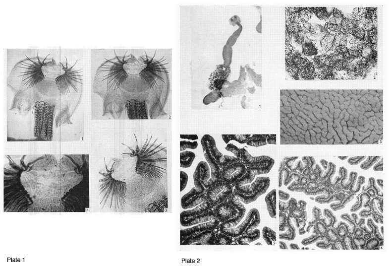 PLATE 1. Echinobothrium acanthinophyllum sp. nov.
Fig. 1. Scolex, lateral view, focused to show apical hooks and peduncle hooks.
Fig. 2. Scolex, lateral view, focused to show the spines on the bothridia, the intrinsic muscles
of the rostellum, the two protractors and the elevator muscles of the apical hooks.
Fig. 3. The two apical groups of hooks. Of the 23 large hooks there is an overlap in two
places on either side at the far end of each row (hooks 2 and 3, 4 and 5). All four small hooks
at the ends of each row are not in focus.
Fig. 4. Apex of scolex showing superficial longitudinal muscles, anterior dorso-ventral
muscles and the circular muscles around the dorsal and ventral lobes of the rostellum. PLATE 2. Echinobothrium acanthinophyllum sp. nov.
Fig. 1. Entire, slightly flattened specimen. Last proglottid, only, gravid.
Fig. 2. Egg clusters in uterus of gravid proglottid.
Fig. 3. Internal surface of spiral valve of Raja montagui Fowler showing primary and
vertical folds.
Fig. 4. Horizontal section through primary folds showing vertical folds on either side, some
joining to form tubes.
Fig. 5. An enlarged portion of the same.
PLATE 1. Echinobothrium acanthinophyllum sp. nov.
Fig. 1. Scolex, lateral view, focused to show apical hooks and peduncle hooks.
Fig. 2. Scolex, lateral view, focused to show the spines on the bothridia, the intrinsic muscles
of the rostellum, the two protractors and the elevator muscles of the apical hooks.
Fig. 3. The two apical groups of hooks. Of the 23 large hooks there is an overlap in two
places on either side at the far end of each row (hooks 2 and 3, 4 and 5). All four small hooks
at the ends of each row are not in focus.
Fig. 4. Apex of scolex showing superficial longitudinal muscles, anterior dorso-ventral
muscles and the circular muscles around the dorsal and ventral lobes of the rostellum. PLATE 2. Echinobothrium acanthinophyllum sp. nov.
Fig. 1. Entire, slightly flattened specimen. Last proglottid, only, gravid.
Fig. 2. Egg clusters in uterus of gravid proglottid.
Fig. 3. Internal surface of spiral valve of Raja montagui Fowler showing primary and
vertical folds.
Fig. 4. Horizontal section through primary folds showing vertical folds on either side, some
joining to form tubes.
Fig. 5. An enlarged portion of the same. 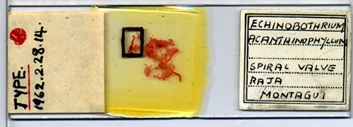

Best viewed in Firefox


