Line Drawing 1

FIGURES 7-11. Ditrachybothridium piliformis n. sp. Segment anatomy. 7. Parasagittal longitudinal section. 8. Transverse section through segment at level indicated by arrow at 8 in Figure 7. 9. T... MoreFIGURES 7-11. Ditrachybothridium piliformis n. sp. Segment anatomy. 7. Parasagittal longitudinal section. 8. Transverse section through segment at level indicated by arrow at 8 in Figure 7. 9. Transverse section through segment at level indicated by arrow at 9 in Figure 7. 10. Transverse section through segment at level indicated by arrow at 10 in Figure 7. Abbreviations: C, cirrus; CP, cirrus pouch; Def, vas deferens; Dev, dorsal exretory vessel; Mh, Mehliss gland; Od, oviduct; Ooc, ootype; Ov, ovary; T, testis; Ut, uterus; Utd, uterine duct; Vev, ventral excretory vessel; Vg, vagina; Vit, vitelline folicles; Vtd, vitelline duct. |
Line Drawing 2

11. Schematic representation of the genital complex, lateral view. Abbreviations: C, cirrus; CP, cirrus pouch; Def, vas deferens; Dev, dorsal exretory vessel; Mh, Mehliss gland; Od, oviduct; Ooc, oo... More11. Schematic representation of the genital complex, lateral view. Abbreviations: C, cirrus; CP, cirrus pouch; Def, vas deferens; Dev, dorsal exretory vessel; Mh, Mehliss gland; Od, oviduct; Ooc, ootype; Ov, ovary; T, testis; Ut, uterus; Utd, uterine duct; Vev, ventral excretory vessel; Vg, vagina; Vit, vitelline folicles; Vtd, vitelline duct. |
Photo Micrograph
|
Scanning Electron Micrograph

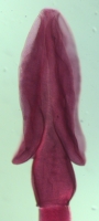
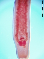
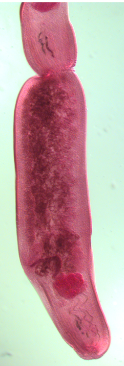
FIGURES 1-6. Ditrachybothridium piliformis n. sp. electron micrographs. 1. Scolex. Bar = 215um. 2. Scolex in cross section, showing median longitudinal furrow (arrow) and dorsal and ventral longit... MoreFIGURES 1-6. Ditrachybothridium piliformis n. sp. electron micrographs. 1. Scolex. Bar = 215um. 2. Scolex in cross section, showing median longitudinal furrow (arrow) and dorsal and ventral longitudinal muscle bundles (arrowhead). Bar = 100um. 3. Lateral view of scolex, showing pectinate spinitriches (arrowhead) and filitriches (*). Bar = 6um. 4. TEM section showing detail of pectinate spinitriches (arrowhead) and filitriches (*). Bar = 0.25um. 5. TEM section showing spatulate spinitriches (arrow) and long filitriches (*) covering proximal surfaces of bothria. Bar = 15um. 6a. Eggs. Bar = 5um. 6b. Detail of egg showing mucron. Bar = 1.25um. bo, bothria; cp, cephalic peduncle; D, dorsal; lm, longitudinal muscle bundle; mu, mucron; V, ventral. |

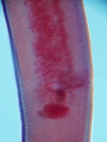
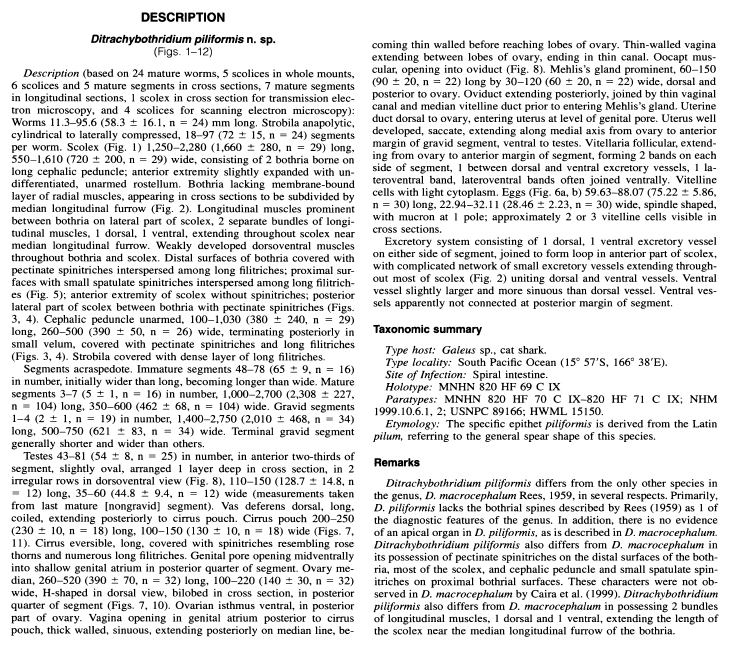
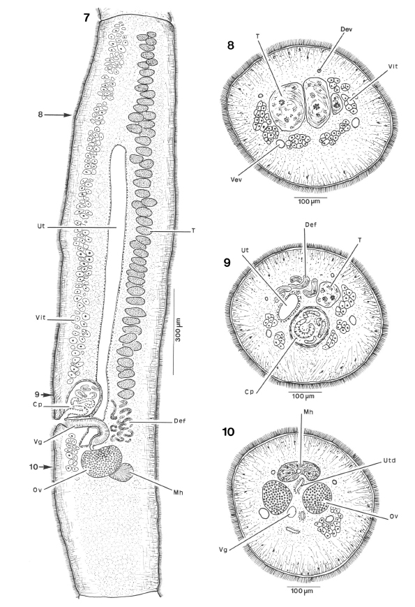 FIGURES 7-11. Ditrachybothridium piliformis n. sp. Segment anatomy. 7. Parasagittal longitudinal section. 8. Transverse section through segment at level indicated by arrow at 8 in Figure 7. 9. Transverse section through segment at level indicated by arrow at 9 in Figure 7. 10. Transverse section through segment at level indicated by arrow at 10 in Figure 7. Abbreviations: C, cirrus; CP, cirrus pouch; Def, vas deferens; Dev, dorsal exretory vessel; Mh, Mehliss gland; Od, oviduct; Ooc, ootype; Ov, ovary; T, testis; Ut, uterus; Utd, uterine duct; Vev, ventral excretory vessel; Vg, vagina; Vit, vitelline folicles; Vtd, vitelline duct.
 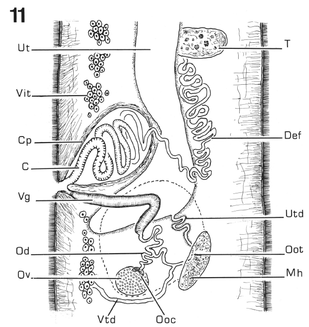 11. Schematic representation of the genital complex, lateral view. Abbreviations: C, cirrus; CP, cirrus pouch; Def, vas deferens; Dev, dorsal exretory vessel; Mh, Mehliss gland; Od, oviduct; Ooc, ootype; Ov, ovary; T, testis; Ut, uterus; Utd, uterine duct; Vev, ventral excretory vessel; Vg, vagina; Vit, vitelline folicles; Vtd, vitelline duct.
 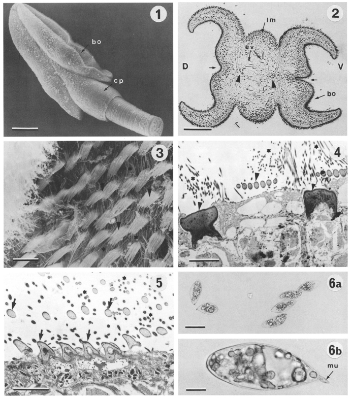 FIGURES 1-6. Ditrachybothridium piliformis n. sp. electron micrographs. 1. Scolex. Bar = 215um. 2. Scolex in cross section, showing median longitudinal furrow (arrow) and dorsal and ventral longitudinal muscle bundles (arrowhead). Bar = 100um. 3. Lateral view of scolex, showing pectinate spinitriches (arrowhead) and filitriches (*). Bar = 6um. 4. TEM section showing detail of pectinate spinitriches (arrowhead) and filitriches (*). Bar = 0.25um. 5. TEM section showing spatulate spinitriches (arrow) and long filitriches (*) covering proximal surfaces of bothria. Bar = 15um. 6a. Eggs. Bar = 5um. 6b. Detail of egg showing mucron. Bar = 1.25um. bo, bothria; cp, cephalic peduncle; D, dorsal; lm, longitudinal muscle bundle; mu, mucron; V, ventral.
  BMNH1999.10.6.1-2 (paratype)
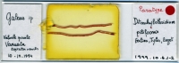
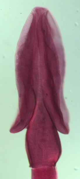
 BMNH1999.10.6.1-2 (paratype)
  BMNH1999.10.6.1-2 (paratype)

