Cestode Scientific Name
| Species ID | 157 |
|---|---|
| Order | Caryophyllidea |
| Family | Capingentidae |
| Subfamily | |
| Genus | Glaridacris |
| Species | hexacotyle |
| Authority | (Linton, 1897) Hunter, 1927 |
| Taxonomic Status | Synonym |
| Valid Name | Isoglaridacris hexacotyle (Linton, 1897) Mackiewicz, 1968 |
| Synonyms | Monobothrium hexacotyle Linton, 1897 Caryophyllaeus hexacotyle (Linton, 1897) Woodland, 1923 |
| Genus Record | No |
| Type Species | No |
| Verified | Yes |
| Verified By | T. Scholz |
| Citation(s) |
Hunter III, G. V. 1927. Notes on the Caryophyllaeidae of North America. Journal of Parasitology 14(1): 16-26. (637) Download PDF |
| Redescription | |
| Scientific Name Notes |
Record Data
| Date (MM/DD/YYYY) | Action | User Name |
|---|---|---|
| 10/13/2005 | Created | Scholz, Oros |
| 07/23/2014 | Modified | |
| 04/02/2020 | Modified | T. Scholz |
| 08/12/2021 | Modified | T. Scholz |
Type Host
| Host Class | |||||||
|---|---|---|---|---|---|---|---|
| Host Order | |||||||
| Host Family | |||||||
|
Type Host (Literal) |
|
||||||
|
Type Host (Valid) |
|
||||||
| Additional Host(s) | |||||||
| Site in Host | |||||||
| Host Notes |
Type Locality
| Country | |
|---|---|
| Body of Water | |
| Island(s) | |
| City/Region | |
| Coordinates | |
| DD Latitude | |
| DD Longitude | |
| Additional Localities | |
| Locality Notes |
Specimens
| Type Material | |
|---|---|
| Total Number of Type Specimens | |
| Voucher Material | |
| Specimen Notes |
Data are given as in original description unless otherwise indicated.
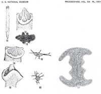
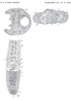
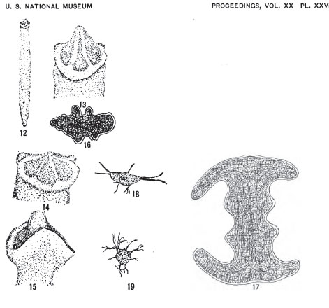 Plate XXVII Fig. 12. Ventral view of specimen. Enlarged three times. Fig. 13. Head of same, side view. Enlarged fifteen times. Fig. 14. Head of another specimen, side view. Enlarged fifteen times. Fig. 15. Marginal view of head. Enlarged fifteen times. Fig. 16. Transverse section near anterior end of head. Zeiss 2/A, draw-tube open. Fig. 17. Transverse section of head farther back than fig. 16. Zeiss 2/A, draw-tube open. Fig. 18. Cell from parenchyma (see Plate XXVIII, Fig. 2). Zeiss 4/D, draw-tube open. Fig. 19. Another cell from same. Zeiss 2/D, draw-tube open.
 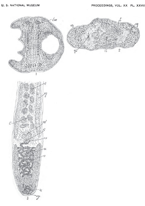 Plate XXVIII Fig. 1. Transverse section toward base of head. Zeiss 2/A, draw-tube open. Fig. 2. Transverse section through middle of body, ge. cell in parenchynea, see Plate XXVII, Figs. 18 and 19. Zeiss 2/A, draw-tube open. Fig. 3. Diagrammatic sketch, ventral view.
 Best viewed in Firefox


