Line Drawing 1

6 Dallarés et al.
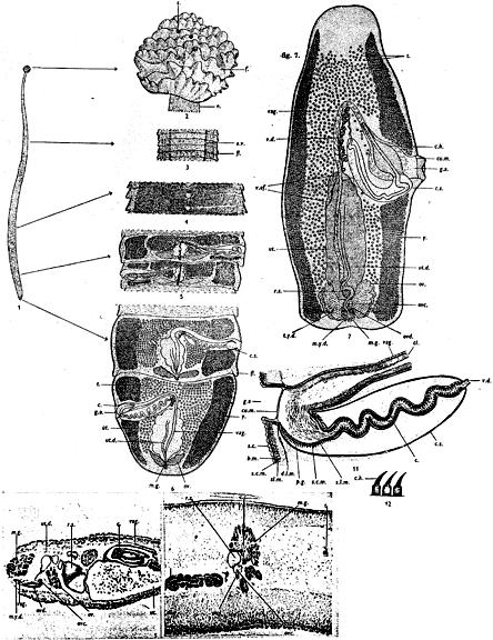
Figure 2. Line drawings of Carrassoniella sinuosiceps. A, scolex. B, outlines of adult worm in convoluted and extended state. C, eggs. D, detail of terminal genitalia. E, late mat... More6 Dallarés et al.
Figure 2. Line drawings of Carrassoniella sinuosiceps. A, scolex. B, outlines of adult worm in convoluted and extended state. C, eggs. D, detail of terminal genitalia. E, late mature proglottid. Abbreviations: c, cirrus; cs, cirrus sac; ex, excretory vessel; mg, Mehlis gland; ov, ovary; t, testis; ut, uterus; va, vagina; vd, vas deferens; vf, vitelline follicle. |
Line Drawing 2
|
Photo Micrograph
|
Scanning Electron Micrograph

Figure 3. Scanning electron micrographs of Carrassoniella sinuosiceps. A, scolex and neck region in dorsoventral view; small letters indicate locations of details in E and F. B, detail of apical part ... MoreFigure 3. Scanning electron micrographs of Carrassoniella sinuosiceps. A, scolex and neck region in dorsoventral view; small letters indicate locations of details in E and F. B, detail of apical part of scolex illustrating accessory suckers; small letters indicate locations of details in C, D, G and H. C, tegumental pore on outer surface of accessory sucker. D, papilliform filitriches on inner surface of accessory sucker. E, detail of scutellate neck. F, capilliform filitriches on strobila. G, gongylate columnar spinitriches on distal bothridial surface. H, trullate spinitriches interspersed with acicular filitriches on proximal bothridial surface. I, external labial structures narrowing genital passage. |


 6 Dallarés et al.
Figure 2. Line drawings of Carrassoniella sinuosiceps. A, scolex. B, outlines of adult worm in convoluted and extended state. C, eggs. D, detail of terminal genitalia. E, late mature proglottid. Abbreviations: c, cirrus; cs, cirrus sac; ex, excretory vessel; mg, Mehlis gland; ov, ovary; t, testis; ut, uterus; va, vagina; vd, vas deferens; vf, vitelline follicle.
6 Dallarés et al.
Figure 2. Line drawings of Carrassoniella sinuosiceps. A, scolex. B, outlines of adult worm in convoluted and extended state. C, eggs. D, detail of terminal genitalia. E, late mature proglottid. Abbreviations: c, cirrus; cs, cirrus sac; ex, excretory vessel; mg, Mehlis gland; ov, ovary; t, testis; ut, uterus; va, vagina; vd, vas deferens; vf, vitelline follicle.  Figure 3. Scanning electron micrographs of Carrassoniella sinuosiceps. A, scolex and neck region in dorsoventral view; small letters indicate locations of details in E and F. B, detail of apical part of scolex illustrating accessory suckers; small letters indicate locations of details in C, D, G and H. C, tegumental pore on outer surface of accessory sucker. D, papilliform filitriches on inner surface of accessory sucker. E, detail of scutellate neck. F, capilliform filitriches on strobila. G, gongylate columnar spinitriches on distal bothridial surface. H, trullate spinitriches interspersed with acicular filitriches on proximal bothridial surface. I, external labial structures narrowing genital passage.


