Line Drawing 1

Figure 1. Bothriocestus cuspidatus (Cooper, 1917) n. comb. from Sander vitreus (Mitchill, 1818). (A) Anterior and middle part of strobila, Fox River, Wisconsin, USA; note craspedote, trapezoidal progl... MoreFigure 1. Bothriocestus cuspidatus (Cooper, 1917) n. comb. from Sander vitreus (Mitchill, 1818). (A) Anterior and middle part of strobila, Fox River, Wisconsin, USA; note craspedote, trapezoidal proglottids, with primary, secondary, and tertiary proglottids differing from each other in size. (B) Anterior end of body and scolex, lateral view. (C) Anterior end of body and scolex, dorsoventral view, Lake Winnipeg, Manitoba, Canada.
Figure 3. Bothriocestus cuspidatus (Cooper, 1917) n. comb. from Sander vitreus (Mitchill, 1818), Lake Winnipeg, Manitoba, Canada (A, C), and Assiniboine River, Manitoba, Canada (B). (A) Mature proglottid, ventral view; median vitelline follicles are not illustrated sinistrally. (B) Dextral side of 2 mature proglottids showing tightly packed testes and nerve cord, ventral view; only lateralmost and few medianmost vitelline follicles are illustrated. (C) Distribution of vitelline follicles in 2 gravid proglottids, dorsal view; only medianmost and lateralmost follicles are illustrated. |
Line Drawing 2
|
Photo Micrograph

Figure 2. Scanning electron micrographs of Bothriocestus cuspidatus (Cooper, 1917) n. comb. from Sander vitreus (Mitchill, 1818), Lake Winnipeg, Manitoba, Canada (AD), and Wolf River, Wisconsin, USA ... MoreFigure 2. Scanning electron micrographs of Bothriocestus cuspidatus (Cooper, 1917) n. comb. from Sander vitreus (Mitchill, 1818), Lake Winnipeg, Manitoba, Canada (AD), and Wolf River, Wisconsin, USA (E). (A) Anterior part, frontal view; note markedly trapeziform proglottids of different sizes. (B) Scolex, lateral view. (C) Scolex, subapical, frontal view. (D) Scolex, apical view; note apical disc. (E) Scolex, frontal view. |
Scanning Electron Micrograph
|
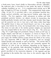


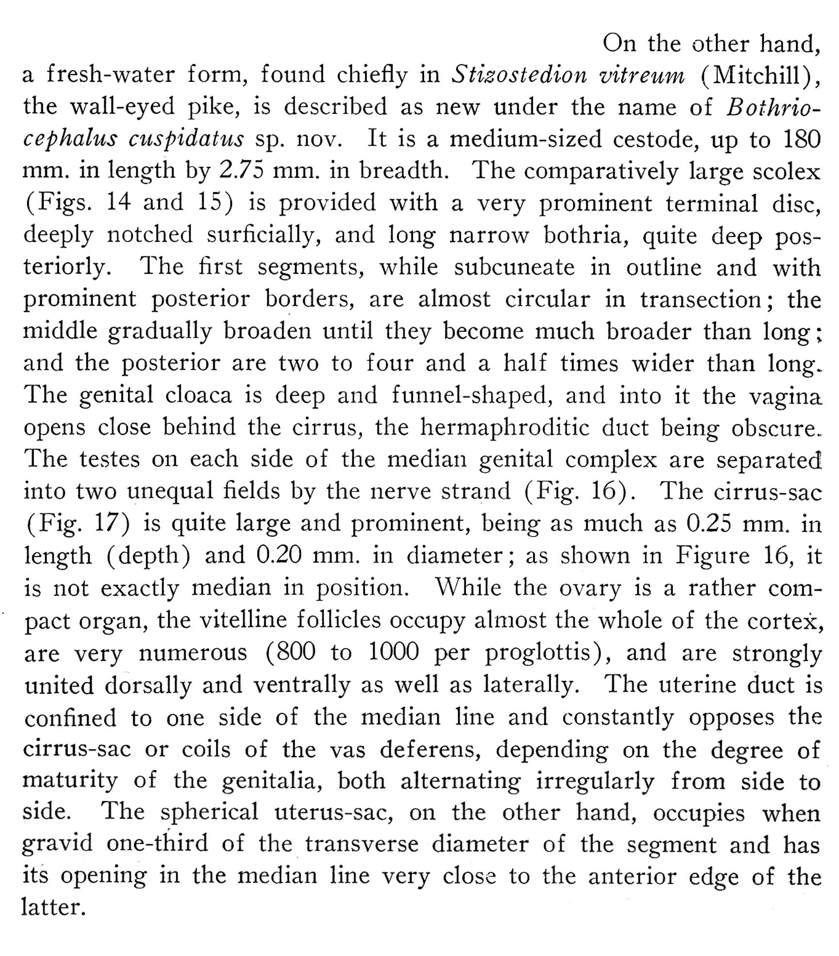
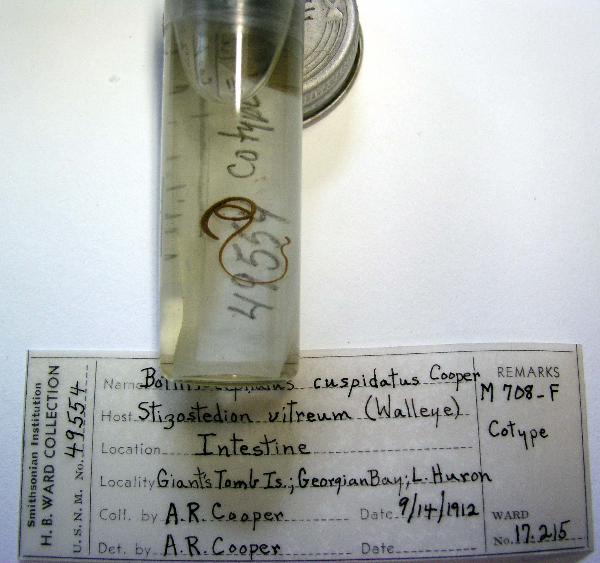
 PLATE II. FIGURES 14-17. Bothriocephalus cuspidatus n. sp. 14. Scolex, surficial view. 15. Scolex, lateral view. 16. Toto of ripe proglottides, posterios in deeper optical section. 17. Median sagittal section, composite.
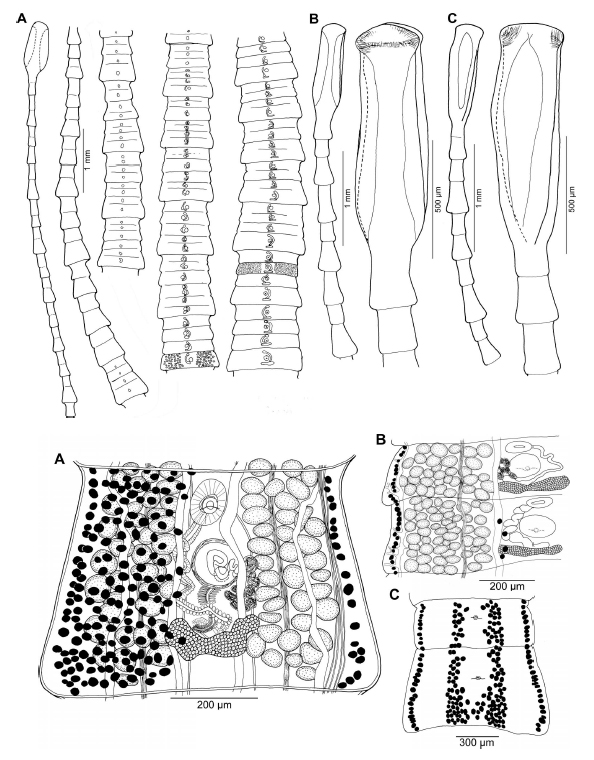 Figure 1. Bothriocestus cuspidatus (Cooper, 1917) n. comb. from Sander vitreus (Mitchill, 1818). (A) Anterior and middle part of strobila, Fox River, Wisconsin, USA; note craspedote, trapezoidal proglottids, with primary, secondary, and tertiary proglottids differing from each other in size. (B) Anterior end of body and scolex, lateral view. (C) Anterior end of body and scolex, dorsoventral view, Lake Winnipeg, Manitoba, Canada.
Figure 3. Bothriocestus cuspidatus (Cooper, 1917) n. comb. from Sander vitreus (Mitchill, 1818), Lake Winnipeg, Manitoba, Canada (A, C), and Assiniboine River, Manitoba, Canada (B). (A) Mature proglottid, ventral view; median vitelline follicles are not illustrated sinistrally. (B) Dextral side of 2 mature proglottids showing tightly packed testes and nerve cord, ventral view; only lateralmost and few medianmost vitelline follicles are illustrated. (C) Distribution of vitelline follicles in 2 gravid proglottids, dorsal view; only medianmost and lateralmost follicles are illustrated.
Figure 1. Bothriocestus cuspidatus (Cooper, 1917) n. comb. from Sander vitreus (Mitchill, 1818). (A) Anterior and middle part of strobila, Fox River, Wisconsin, USA; note craspedote, trapezoidal proglottids, with primary, secondary, and tertiary proglottids differing from each other in size. (B) Anterior end of body and scolex, lateral view. (C) Anterior end of body and scolex, dorsoventral view, Lake Winnipeg, Manitoba, Canada.
Figure 3. Bothriocestus cuspidatus (Cooper, 1917) n. comb. from Sander vitreus (Mitchill, 1818), Lake Winnipeg, Manitoba, Canada (A, C), and Assiniboine River, Manitoba, Canada (B). (A) Mature proglottid, ventral view; median vitelline follicles are not illustrated sinistrally. (B) Dextral side of 2 mature proglottids showing tightly packed testes and nerve cord, ventral view; only lateralmost and few medianmost vitelline follicles are illustrated. (C) Distribution of vitelline follicles in 2 gravid proglottids, dorsal view; only medianmost and lateralmost follicles are illustrated. 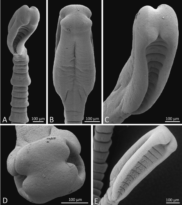 Figure 2. Scanning electron micrographs of Bothriocestus cuspidatus (Cooper, 1917) n. comb. from Sander vitreus (Mitchill, 1818), Lake Winnipeg, Manitoba, Canada (AD), and Wolf River, Wisconsin, USA (E). (A) Anterior part, frontal view; note markedly trapeziform proglottids of different sizes. (B) Scolex, lateral view. (C) Scolex, subapical, frontal view. (D) Scolex, apical view; note apical disc. (E) Scolex, frontal view.