Line Drawing 1
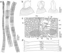
Line drawings of Diphyllobothrium sprakeri n. sp. from Zalophus californianus, California, USA. a Selected anterior, middle and posterior proglottids of holotype, ventral view. b Scoleces, dorsovent... MoreLine drawings of Diphyllobothrium sprakeri n. sp. from Zalophus californianus, California, USA. a Selected anterior, middle and posterior proglottids of holotype, ventral view. b Scoleces, dorsoventral view. Scolex of holotype marked with an asterisk. c Genitalia of mature proglottids of holotype, ventral view, vitelline follicles omitted on left side and testes on the right side of proglottid. d Schematic drawing of gravid proglottids of holotype showing the position of the sac-like structure of the uterus. Abbreviations: cs, cirrus sac; esv, external seminal vesicle; ga, genital atrium; mg, Mehlis gland; ov, ovary; te, testes; up, uterine pore; uss, uterine sac-like structure; ut, uterus; vf, vitelline follicle; vg, vagina. |
Line Drawing 2
|
Photo Micrograph
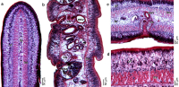
Photomicrographs of histological sections of holotype of Diphyllobothrium sprakeri n. sp. from Zalophus californianus, California, USA. a Gravid proglottid, cross section. b Gravid proglottids, sagi... MorePhotomicrographs of histological sections of holotype of Diphyllobothrium sprakeri n. sp. from Zalophus californianus, California, USA. a Gravid proglottid, cross section. b Gravid proglottids, sagittal section. c Detail of cirrus sac and genital atrium in gravid proglottid, cross section. d Detail of longitudinal musculature. Abbreviations: cs, cirrus sac; esv, external seminal vesicle; lm, longitudinal musculature; te, testes; uss, uterine sac-like structure; vf, vitelline follicle. |
Scanning Electron Micrograph

Scanning electron micrographs of Diphyllobothrium sprakeri n. sp. from Zalophus californianus, California, USA. a Capilliform filitriches on the lateral scolex surface. b Coniform spinitriches on th... MoreScanning electron micrographs of Diphyllobothrium sprakeri n. sp. from Zalophus californianus, California, USA. a Capilliform filitriches on the lateral scolex surface. b Coniform spinitriches on the dorsoventral scolex surface. ce Scoleces, dorsoventral view. f Scolex in apical view. g Ventral surface of gravid proglottids showing numerous papillae surrounding the genital atrium. hi Eggs and surface of egg shell. j Detail of egg surface.
with pits |
 Line drawings of Diphyllobothrium sprakeri n. sp. from Zalophus californianus, California, USA. a Selected anterior, middle and posterior proglottids of holotype, ventral view. b Scoleces, dorsoventral view. Scolex of holotype marked with an asterisk. c Genitalia of mature proglottids of holotype, ventral view, vitelline follicles omitted on left side and testes on the right side of proglottid. d Schematic drawing of gravid proglottids of holotype showing the position of the sac-like structure of the uterus. Abbreviations: cs, cirrus sac; esv, external seminal vesicle; ga, genital atrium; mg, Mehlis gland; ov, ovary; te, testes; up, uterine pore; uss, uterine sac-like structure; ut, uterus; vf, vitelline follicle; vg, vagina.
 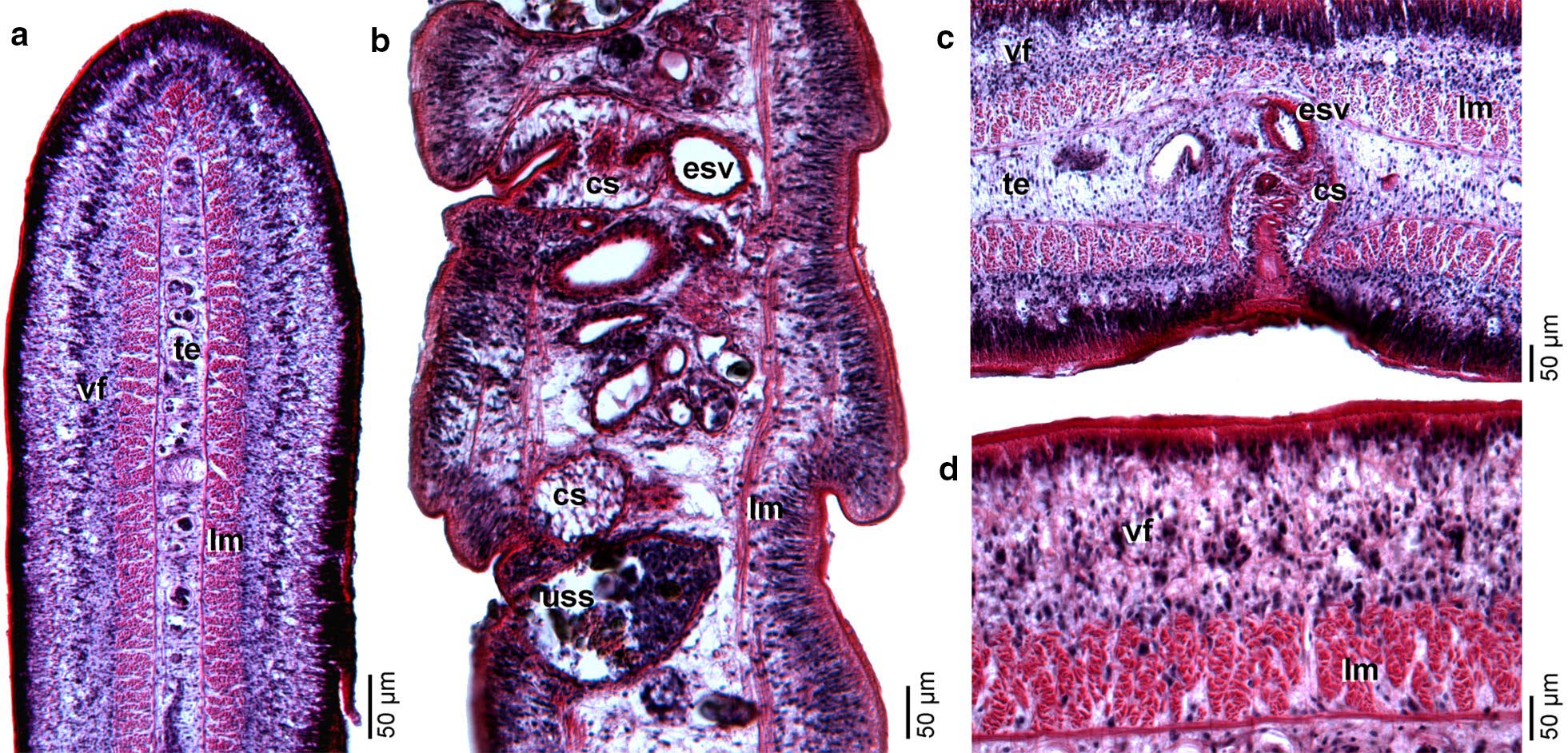 Photomicrographs of histological sections of holotype of Diphyllobothrium sprakeri n. sp. from Zalophus californianus, California, USA. a Gravid proglottid, cross section. b Gravid proglottids, sagittal section. c Detail of cirrus sac and genital atrium in gravid proglottid, cross section. d Detail of longitudinal musculature. Abbreviations: cs, cirrus sac; esv, external seminal vesicle; lm, longitudinal musculature; te, testes; uss, uterine sac-like structure; vf, vitelline follicle.
 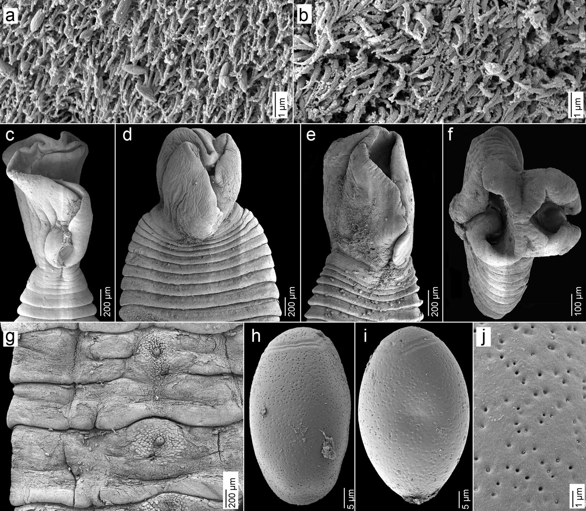 Scanning electron micrographs of Diphyllobothrium sprakeri n. sp. from Zalophus californianus, California, USA. a Capilliform filitriches on the lateral scolex surface. b Coniform spinitriches on the dorsoventral scolex surface. ce Scoleces, dorsoventral view. f Scolex in apical view. g Ventral surface of gravid proglottids showing numerous papillae surrounding the genital atrium. hi Eggs and surface of egg shell. j Detail of egg surface.
with pits
Scanning electron micrographs of Diphyllobothrium sprakeri n. sp. from Zalophus californianus, California, USA. a Capilliform filitriches on the lateral scolex surface. b Coniform spinitriches on the dorsoventral scolex surface. ce Scoleces, dorsoventral view. f Scolex in apical view. g Ventral surface of gravid proglottids showing numerous papillae surrounding the genital atrium. hi Eggs and surface of egg shell. j Detail of egg surface.
with pits 

