Line Drawing 1
From Herzog & Jensen, 2022 (Cit# 7426). Figure 8 Line drawings of Rhinoptericola jensenae (Schaeffner & Beveridge, 2012b) n. comb. (A) Whole worm (QM G239457; voucher). (B) Scolex (QM G239461; voucher... MoreFrom Herzog & Jensen, 2022 (Cit# 7426). Figure 8 Line drawings of Rhinoptericola jensenae (Schaeffner & Beveridge, 2012b) n. comb. (A) Whole worm (QM G239457; voucher). (B) Scolex (QM G239461; voucher). (C) Terminal proglottid (QM G239460; voucher); circumcortical vitelline follicles are drawn only on the lateral margins and in the region delimited by dashed lines. Full-size image DOI: 10.7717/peerj.12865/fig-8 |
Line Drawing 2

From Herzog & Jensen, 2022 (Cit# 7426). Figure 9 Line drawings of the tentacular armature of Rhinoptericola jensenae (Schaeffner & Beveridge, 2012b) n. comb. (A) Metabasal armature, bothrial surface (... MoreFrom Herzog & Jensen, 2022 (Cit# 7426). Figure 9 Line drawings of the tentacular armature of Rhinoptericola jensenae (Schaeffner & Beveridge, 2012b) n. comb. (A) Metabasal armature, bothrial surface (USNM 1661573; voucher). (B) Metabasal armature, antibothrial surface (USNM 1661573; voucher). (C) Metabasal armature, distal antibothrial surface, showing a reduction to six hooks per principal row (LRP 10574; voucher). (D) Basal armature, bothrial surface (QM G239461; voucher). (E) Basal armature, antibothrial surface (QM G239461; voucher). (F) Comparison of metabasal hook shapes. Full-size image DOI: 10.7717/peerj.12865/fig-9 |
Photo Micrograph
|
Scanning Electron Micrograph
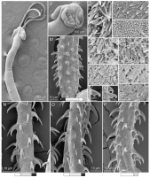
From Herzog & Jensen, 2022 (Cit# 7426). Figure 10 Scanning electron micrographs of Rhinoptericola jensenae (Schaeffner & Beveridge, 2012b) n. comb. (A) Scolex; small letters indicate the location of d... MoreFrom Herzog & Jensen, 2022 (Cit# 7426). Figure 10 Scanning electron micrographs of Rhinoptericola jensenae (Schaeffner & Beveridge, 2012b) n. comb. (A) Scolex; small letters indicate the location of details shown in (HI). (B) Bothria; small letters indicate the location of details shown in (CG). (C) Distal bothrial surface. (D) Proximal bothrial surface near the bothrial rim. (E) Proximal bothria surface away from the bothrial rim. (F) Surface of the scolex proper between the bothria. (G) Surface of the scolex proper at the apex. (H) Surface of the pars vaginalis.
(I) Surface of the pars bulbosa. (J) Strobilar surface. (K) and (L) Falcate, erect, dorsoventrally flattened billhooks with short forward protrusions on their lower surface and mucronate tips (i.e., can openershaped billhooks) on the antibothrial surface of the basal armature. (M) Basal armature, antibothrial surface. (N) Metabasal armature, bothrial surface. (O) Metabasal armature, antibothrial surface. (P) Metabasal armature, internal surface. Full-size image DOI: 10.7717/peerj.12865/fig-10 |
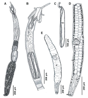
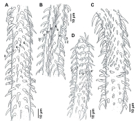
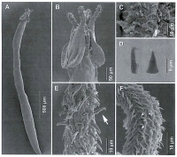
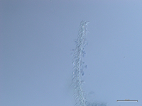
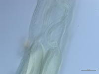

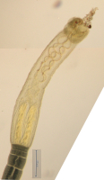
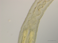
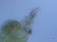
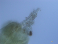
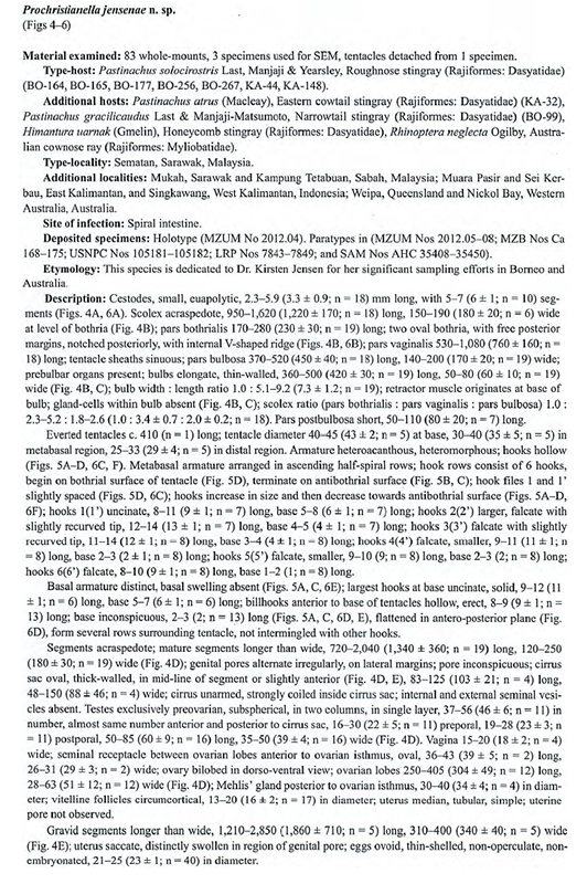
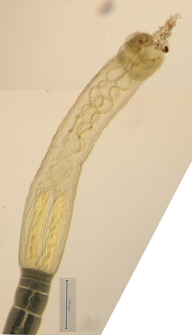
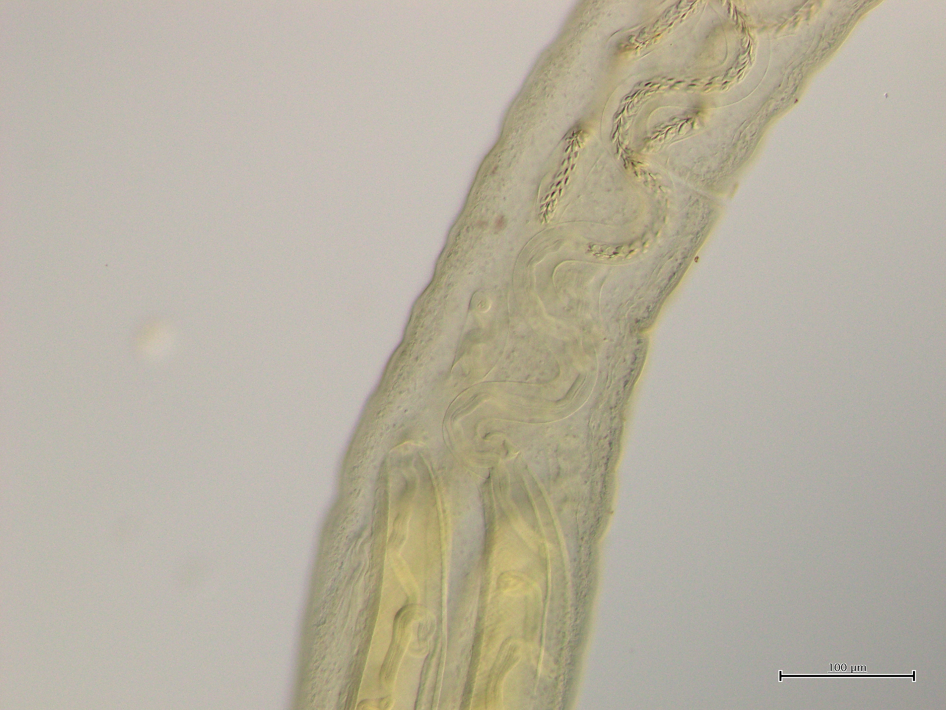
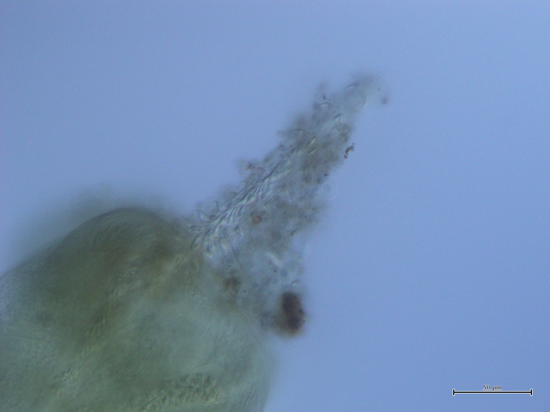
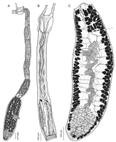
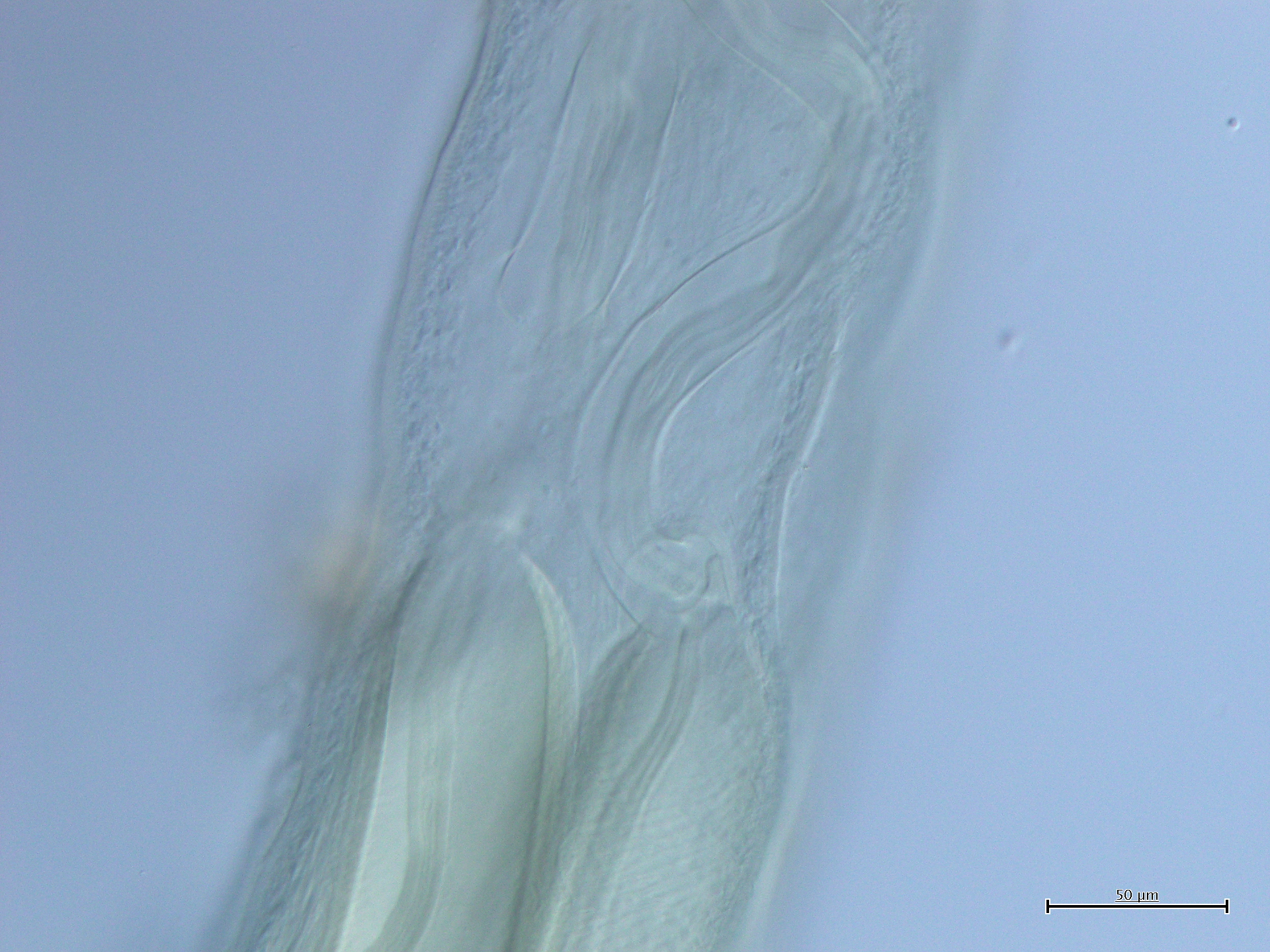
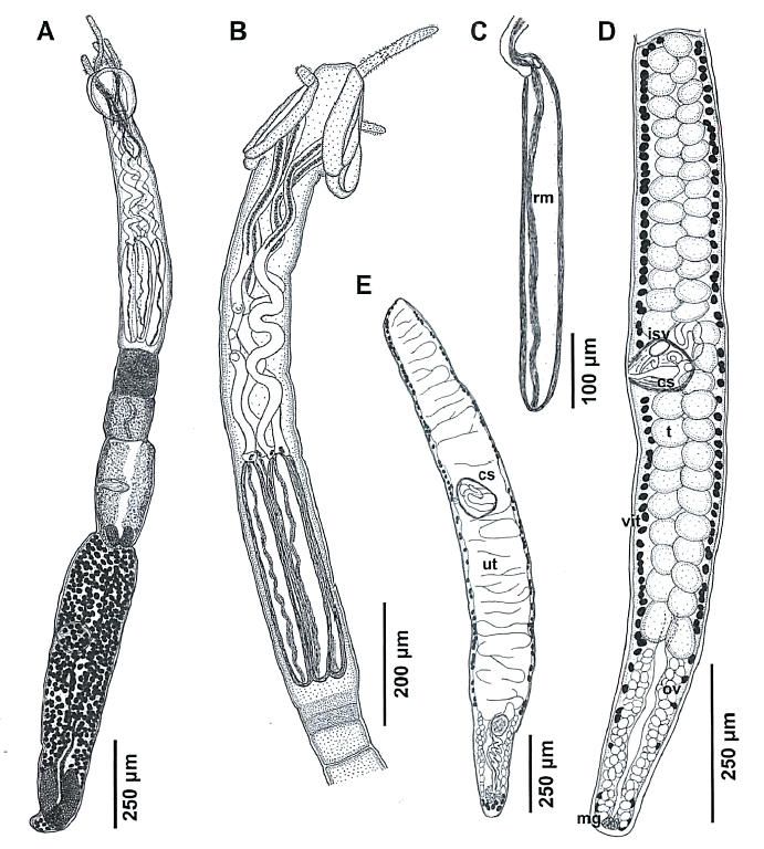 FIGURE 4. Prochristianella jensenae n. sp. from Pastinachus solocirostris A. Complete specimen. B. Scolex. C. Bulb; note absence of gland-cells. D. Mature segment. E. Gravid segment. Abbreviations: cs, cirrus sac; isv, internal seminal vesicle; mg, Mehlis' gland; ov, ovary; rm, retractor muscle; t, testis; ut, uterus; vit, vitelline follicle.
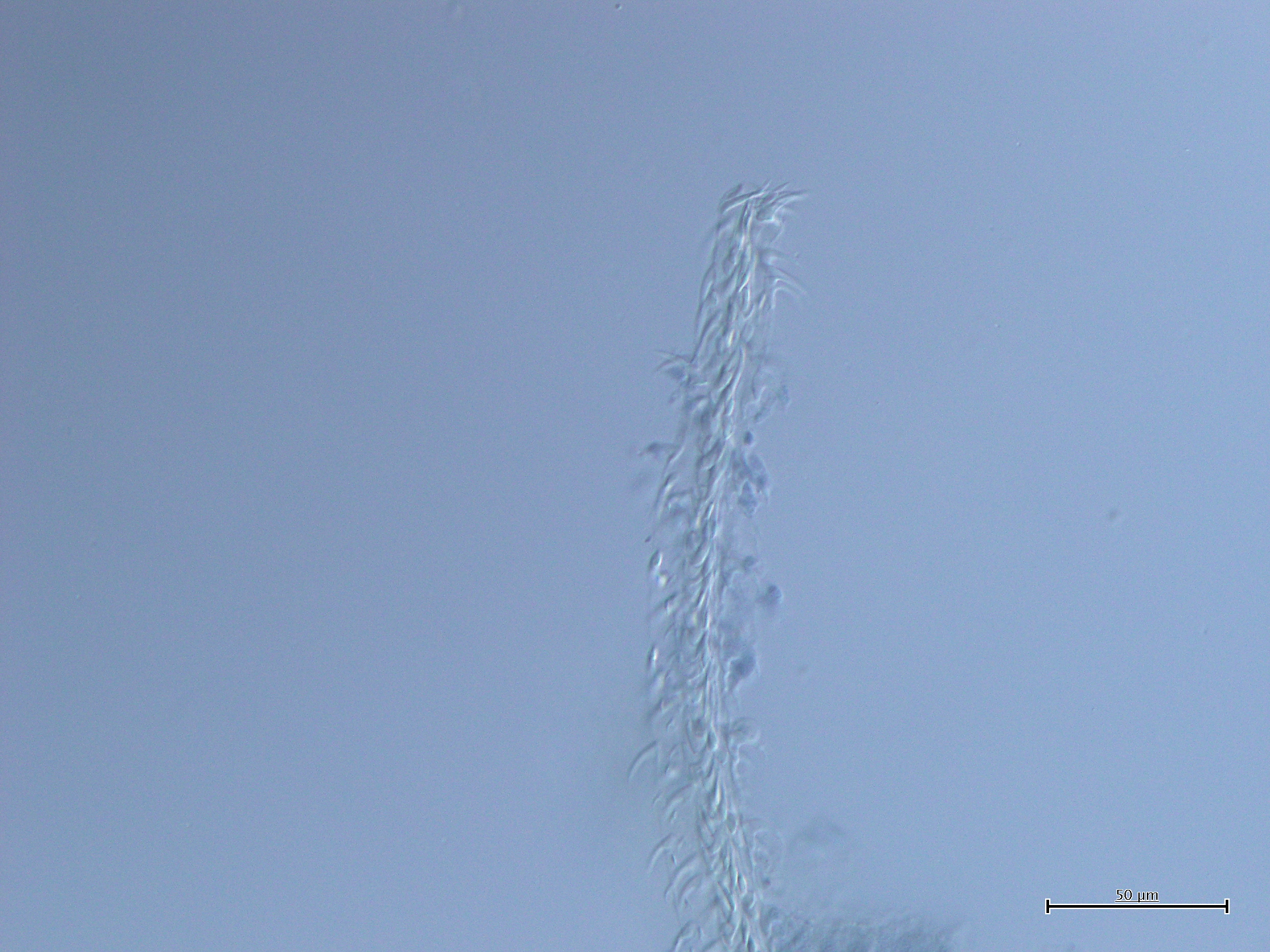
 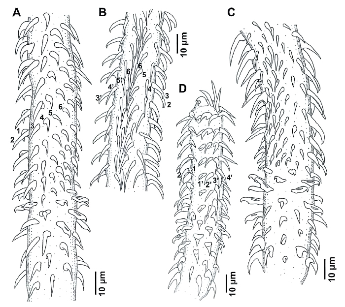 FIGURE 5. Prochristianella jensenae n. sp. from Pastinachus gracilicaudus (A, D) and P. solocirostris (B, C). Tentacular armature. A. Basal and metabasal regions, internal surface. B. Metabasal region, antibothrial surface. C. Basal and metabasal regions, antibothrial surface. D. Basal and metabasal regions, bothrial surface.
 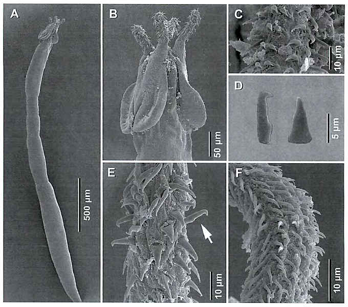 FIGURE 6. Prochristianella jensenae n. sp. from Pastinachus solcirostris. Scanning electron micrographs. A. Complete specimen. B. Scolex in lateral view. C. Tentacular armature; metabasal region, bothrial surface. D. Billhooks in lateral (left) and dorso-ventral (right) views. E. Tentacular armature, basal region, internal surface; note erect billhook. F. Tentacular armature, metabasal region, external surface.
  From Herzog & Jensen, 2022 (Cit# 7426). Figure 8 Line drawings of Rhinoptericola jensenae (Schaeffner & Beveridge, 2012b) n. comb. (A) Whole worm (QM G239457; voucher). (B) Scolex (QM G239461; voucher). (C) Terminal proglottid (QM G239460; voucher); circumcortical vitelline follicles are drawn only on the lateral margins and in the region delimited by dashed lines. Full-size image DOI: 10.7717/peerj.12865/fig-8
 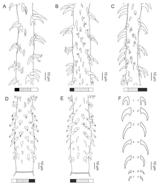 From Herzog & Jensen, 2022 (Cit# 7426). Figure 9 Line drawings of the tentacular armature of Rhinoptericola jensenae (Schaeffner & Beveridge, 2012b) n. comb. (A) Metabasal armature, bothrial surface (USNM 1661573; voucher). (B) Metabasal armature, antibothrial surface (USNM 1661573; voucher). (C) Metabasal armature, distal antibothrial surface, showing a reduction to six hooks per principal row (LRP 10574; voucher). (D) Basal armature, bothrial surface (QM G239461; voucher). (E) Basal armature, antibothrial surface (QM G239461; voucher). (F) Comparison of metabasal hook shapes. Full-size image DOI: 10.7717/peerj.12865/fig-9
  From Herzog & Jensen, 2022 (Cit# 7426). Figure 10 Scanning electron micrographs of Rhinoptericola jensenae (Schaeffner & Beveridge, 2012b) n. comb. (A) Scolex; small letters indicate the location of details shown in (HI). (B) Bothria; small letters indicate the location of details shown in (CG). (C) Distal bothrial surface. (D) Proximal bothrial surface near the bothrial rim. (E) Proximal bothria surface away from the bothrial rim. (F) Surface of the scolex proper between the bothria. (G) Surface of the scolex proper at the apex. (H) Surface of the pars vaginalis.
(I) Surface of the pars bulbosa. (J) Strobilar surface. (K) and (L) Falcate, erect, dorsoventrally flattened billhooks with short forward protrusions on their lower surface and mucronate tips (i.e., can openershaped billhooks) on the antibothrial surface of the basal armature. (M) Basal armature, antibothrial surface. (N) Metabasal armature, bothrial surface. (O) Metabasal armature, antibothrial surface. (P) Metabasal armature, internal surface. Full-size image DOI: 10.7717/peerj.12865/fig-10
From Herzog & Jensen, 2022 (Cit# 7426). Figure 10 Scanning electron micrographs of Rhinoptericola jensenae (Schaeffner & Beveridge, 2012b) n. comb. (A) Scolex; small letters indicate the location of details shown in (HI). (B) Bothria; small letters indicate the location of details shown in (CG). (C) Distal bothrial surface. (D) Proximal bothrial surface near the bothrial rim. (E) Proximal bothria surface away from the bothrial rim. (F) Surface of the scolex proper between the bothria. (G) Surface of the scolex proper at the apex. (H) Surface of the pars vaginalis.
(I) Surface of the pars bulbosa. (J) Strobilar surface. (K) and (L) Falcate, erect, dorsoventrally flattened billhooks with short forward protrusions on their lower surface and mucronate tips (i.e., can openershaped billhooks) on the antibothrial surface of the basal armature. (M) Basal armature, antibothrial surface. (N) Metabasal armature, bothrial surface. (O) Metabasal armature, antibothrial surface. (P) Metabasal armature, internal surface. Full-size image DOI: 10.7717/peerj.12865/fig-10 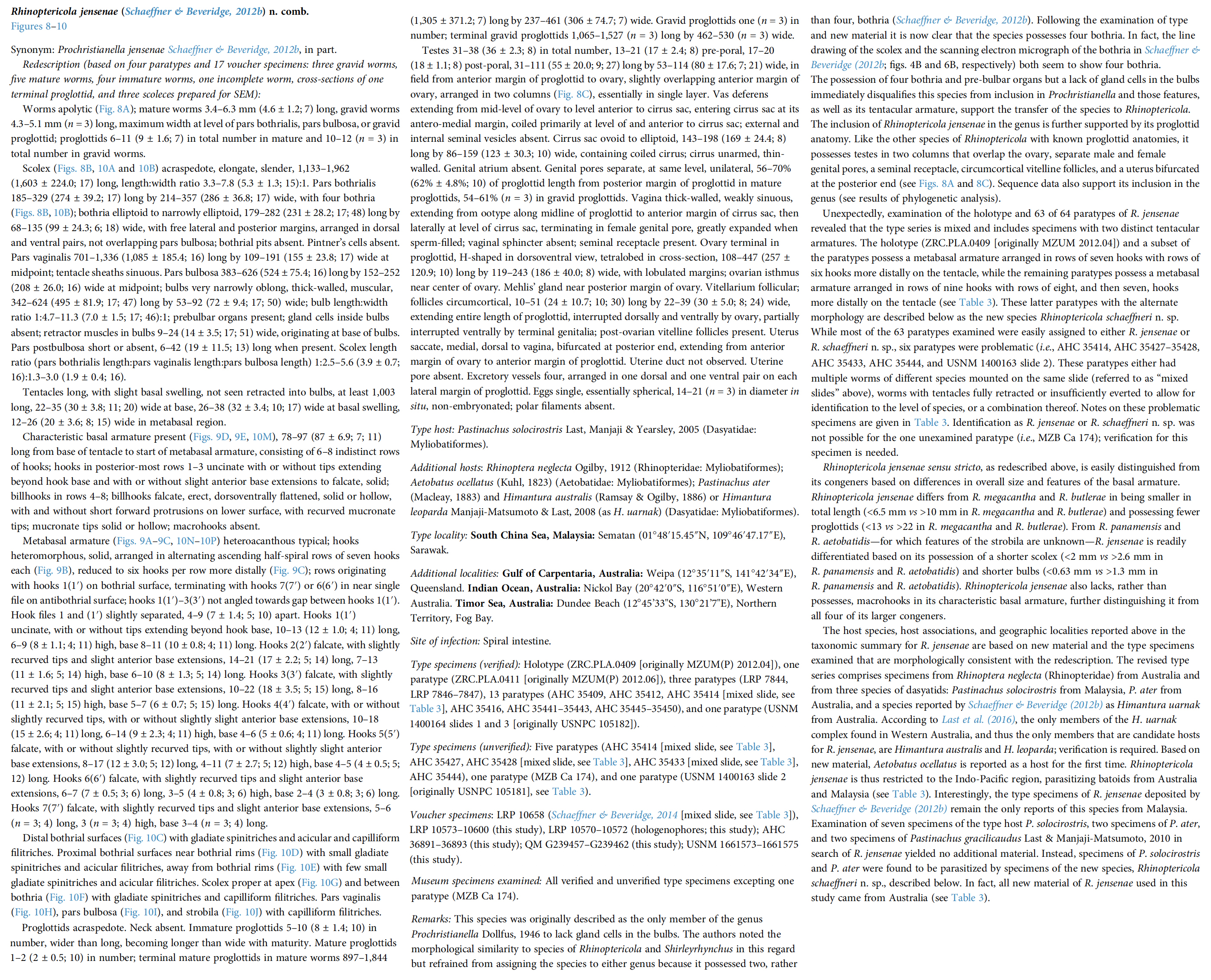
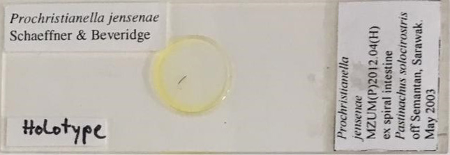 MZUM(P) 2012.04 = ZRC.PLA.0409
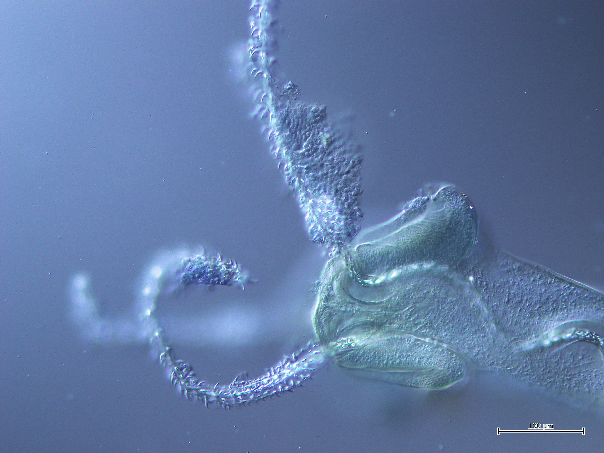
 MZUM(P) 2012.06 = ZRC.PLA.0411