Line Drawing 1

Fig. 2. Line drawings of Biacetabulum isaureae n. sp. from Moxostoma collapsum, South Carolina, USA (US 275d, IPCAS C-591/3). (A) Total view, (B) anterior part with scolex, and (C) posterior part, ven... MoreFig. 2. Line drawings of Biacetabulum isaureae n. sp. from Moxostoma collapsum, South Carolina, USA (US 275d, IPCAS C-591/3). (A) Total view, (B) anterior part with scolex, and (C) posterior part, ventral view. cs, cirrus sac; esv, external seminal vesicle; fgp, female genital pore; mg, Mehlis glands; mgp, male genital pore; ov, ovary; rs, seminal receptacle; te, testes; ut, uterus; vd, vas deferens; vf, vitelline follicles; vtd, vitelline duct. |
Line Drawing 2
|
Photo Micrograph
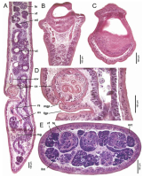
Fig. 3. Histological sections of Biacetabulum isaureae n. sp. from Moxostoma collapsum (US 275d), South Carolina, USA. (A) Sagittal section of the posterior part of body, (B, C) longitudinal sections ... MoreFig. 3. Histological sections of Biacetabulum isaureae n. sp. from Moxostoma collapsum (US 275d), South Carolina, USA. (A) Sagittal section of the posterior part of body, (B, C) longitudinal sections of scolex, and (D) sagittal section of genital pores; E, cross-section of middle portion of body. cs, cirrus-sac; esv, external seminal vesicle; fgp, female genital pore; ilm, internal longitudinal muscles; mg, Mehlis glands; mgp, male genital pore; orc, osmoregulatory canals; ov, ovary; rs, seminal receptacle; te, testes; ut, uterus; vd, vas deferens; vf, vitelline follicles. |
Scanning Electron Micrograph
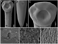
Fig. 4. Scanning electron micrographs of Biacetabulum isaureae n. sp. from Moxostoma collapsum, South Carolina, USA (US 272b, US 275d, IPCAS C-591/3). (A) Anterior part with scolex, (B) posterior part... MoreFig. 4. Scanning electron micrographs of Biacetabulum isaureae n. sp. from Moxostoma collapsum, South Carolina, USA (US 272b, US 275d, IPCAS C-591/3). (A) Anterior part with scolex, (B) posterior part with genital pores, (C) scolex, (D) detail of common genital atrium, (E) microtriches surrounding the common genital atrium, and (F) microtriches on the body. Small letters in A and B indicate position of E and F. |
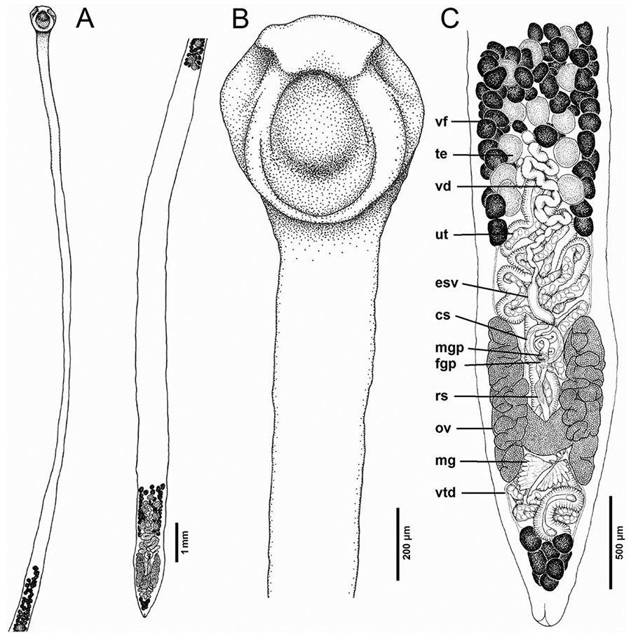 Fig. 2. Line drawings of Biacetabulum isaureae n. sp. from Moxostoma collapsum, South Carolina, USA (US 275d, IPCAS C-591/3). (A) Total view, (B) anterior part with scolex, and (C) posterior part, ventral view. cs, cirrus sac; esv, external seminal vesicle; fgp, female genital pore; mg, Mehlis glands; mgp, male genital pore; ov, ovary; rs, seminal receptacle; te, testes; ut, uterus; vd, vas deferens; vf, vitelline follicles; vtd, vitelline duct.
 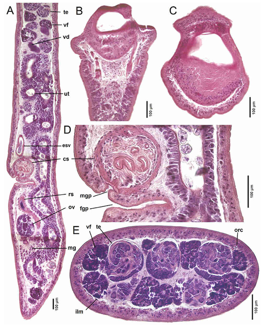 Fig. 3. Histological sections of Biacetabulum isaureae n. sp. from Moxostoma collapsum (US 275d), South Carolina, USA. (A) Sagittal section of the posterior part of body, (B, C) longitudinal sections of scolex, and (D) sagittal section of genital pores; E, cross-section of middle portion of body. cs, cirrus-sac; esv, external seminal vesicle; fgp, female genital pore; ilm, internal longitudinal muscles; mg, Mehlis glands; mgp, male genital pore; orc, osmoregulatory canals; ov, ovary; rs, seminal receptacle; te, testes; ut, uterus; vd, vas deferens; vf, vitelline follicles.
  Fig. 4. Scanning electron micrographs of Biacetabulum isaureae n. sp. from Moxostoma collapsum, South Carolina, USA (US 272b, US 275d, IPCAS C-591/3). (A) Anterior part with scolex, (B) posterior part with genital pores, (C) scolex, (D) detail of common genital atrium, (E) microtriches surrounding the common genital atrium, and (F) microtriches on the body. Small letters in A and B indicate position of E and F.

