Line Drawing 1
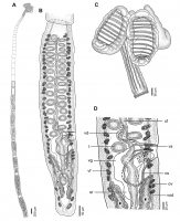
FIGURE 1. Line drawings of Scalithrium kirchneri sp. nov. from Rioraja agassizii. A. entire mature worm (holotype MACNPa 757); B. terminal mature proglottid (paratype MACN-Pa 758/1); C. scolex (paraty... MoreFIGURE 1. Line drawings of Scalithrium kirchneri sp. nov. from Rioraja agassizii. A. entire mature worm (holotype MACNPa 757); B. terminal mature proglottid (paratype MACN-Pa 758/1); C. scolex (paratype MACN-Pa 761); D. detail of terminal genitalia in a terminal mature proglottid (paratype MACN-Pa 758/1). Abbreviations: cs, cirrus sac; ov, ovary; sr, seminal receptacle; t, testes; ut, uterus; vd, vas deferens; vf, vitelline follicle; vg, vagina; vod, ventral osmoregulatory duct; vs, vaginal sphincter. |
Line Drawing 2
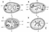
FIGURE 3. Cross sections of terminal mature proglottid of Scalithrium kirchneri sp. nov. from Rioraja agassizii (paratype MACN-Pa 758/13). A. cross section at the level of testes; B. cross section at ... MoreFIGURE 3. Cross sections of terminal mature proglottid of Scalithrium kirchneri sp. nov. from Rioraja agassizii (paratype MACN-Pa 758/13). A. cross section at the level of testes; B. cross section at the level of the genital pore; C. cross section at the level of the seminal receptacle; D. cross section at the level of the ovarian isthmus. Abbreviations: dod, dorsal osmoregulatory duct; ov, ovary; sr, seminal receptacle; t, testes; ut, uterus; vd, vas deferens; vf, vitelline follicle; vg, vagina; vod, ventral osmoregulatory duct; vs, vaginal sphincter. |
Photo Micrograph
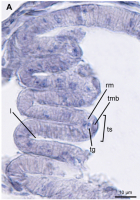
FIGURE 4. Longitudinal sections of bothridium showing the septal musculature. A. Scalithrium kirchneri sp. nov. from Rioraja agassizii (paratype MACN-Pa 759). Abbreviations: l, loculus; rm, radial mus... MoreFIGURE 4. Longitudinal sections of bothridium showing the septal musculature. A. Scalithrium kirchneri sp. nov. from Rioraja agassizii (paratype MACN-Pa 759). Abbreviations: l, loculus; rm, radial muscle; tg, triangular gap; tmb, transverse muscle bundles; ts, transverse septum. |
Scanning Electron Micrograph

FIGURE 2. Scanning electron micrographs of Scalithrium kirchneri sp. nov. from Rioraja agassizii. A. scolex, small letters indicate the location of details shown in figures 2CF; B. surface of scolex ... MoreFIGURE 2. Scanning electron micrographs of Scalithrium kirchneri sp. nov. from Rioraja agassizii. A. scolex, small letters indicate the location of details shown in figures 2CF; B. surface of scolex apex; C. distal bothridial surface; D. proximal bothridial surface; E. surface of stalk; F. surface of cephalic peduncle; G. surface of terminal mature proglottid. |
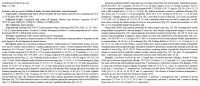
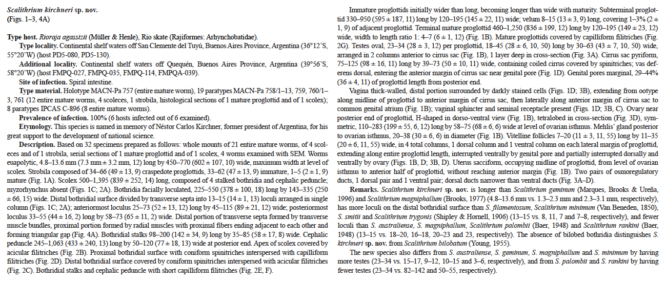
 FIGURE 1. Line drawings of Scalithrium kirchneri sp. nov. from Rioraja agassizii. A. entire mature worm (holotype MACNPa 757); B. terminal mature proglottid (paratype MACN-Pa 758/1); C. scolex (paratype MACN-Pa 761); D. detail of terminal genitalia in a terminal mature proglottid (paratype MACN-Pa 758/1). Abbreviations: cs, cirrus sac; ov, ovary; sr, seminal receptacle; t, testes; ut, uterus; vd, vas deferens; vf, vitelline follicle; vg, vagina; vod, ventral osmoregulatory duct; vs, vaginal sphincter.
  FIGURE 3. Cross sections of terminal mature proglottid of Scalithrium kirchneri sp. nov. from Rioraja agassizii (paratype MACN-Pa 758/13). A. cross section at the level of testes; B. cross section at the level of the genital pore; C. cross section at the level of the seminal receptacle; D. cross section at the level of the ovarian isthmus. Abbreviations: dod, dorsal osmoregulatory duct; ov, ovary; sr, seminal receptacle; t, testes; ut, uterus; vd, vas deferens; vf, vitelline follicle; vg, vagina; vod, ventral osmoregulatory duct; vs, vaginal sphincter.
 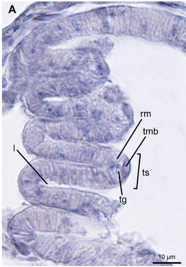 FIGURE 4. Longitudinal sections of bothridium showing the septal musculature. A. Scalithrium kirchneri sp. nov. from Rioraja agassizii (paratype MACN-Pa 759). Abbreviations: l, loculus; rm, radial muscle; tg, triangular gap; tmb, transverse muscle bundles; ts, transverse septum.
  FIGURE 2. Scanning electron micrographs of Scalithrium kirchneri sp. nov. from Rioraja agassizii. A. scolex, small letters indicate the location of details shown in figures 2CF; B. surface of scolex apex; C. distal bothridial surface; D. proximal bothridial surface; E. surface of stalk; F. surface of cephalic peduncle; G. surface of terminal mature proglottid.

