Line Drawing 1
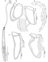
Fig. 4. Guidus magellanicus sp. n. from Bathyraja magellanica (Philippi), line drawings. A entire gravid worm (holotype MACN-Pa No. 747); B scolex (holotype MACN-Pa No. 747); C cocoon; D bothr... MoreFig. 4. Guidus magellanicus sp. n. from Bathyraja magellanica (Philippi), line drawings. A entire gravid worm (holotype MACN-Pa No. 747); B scolex (holotype MACN-Pa No. 747); C cocoon; D bothridium, muscular sphincter relaxed (paratype MACN-Pa No. 748/2); E bothridium, muscular sphincter contracted (paratype IPCAS No. C-888) |
Line Drawing 2
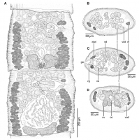
Fig. 5. Guidus magellanicus sp. n. from Bathyraja magellanica (Philippi), line drawings. A subterminal portion of strobila including mature and gravid proglottids, dorsal view (paratype MACN-Pa No. ... MoreFig. 5. Guidus magellanicus sp. n. from Bathyraja magellanica (Philippi), line drawings. A subterminal portion of strobila including mature and gravid proglottids, dorsal view (paratype MACN-Pa No. 748/1); B cross section of mature proglottid at level of testes anterior to cirrus sac; C cross section of mature proglottid at level of genital atrium; D cross section of mature proglottid at level of ovarian isthmus. Abbreviations: cs cirrus sac, dod dorsal osmoregulatory duct, ga genital atrium, oc ovicapt, ov ovary, t testis, vd vas deferens, vf vitelline follicle, vg vagina, vod ventral osmoregulatory duct |
Photo Micrograph
|
Scanning Electron Micrograph
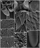
Fig. 6. Guidus magellanicus sp. n. from Bathyraja magellanica (Philippi), scanning electron micrographs. A scolex, small letters indicate locations of detail shown in CE and H, arrow indicates marg... MoreFig. 6. Guidus magellanicus sp. n. from Bathyraja magellanica (Philippi), scanning electron micrographs. A scolex, small letters indicate locations of detail shown in CE and H, arrow indicates marginal indentation; B partial apical view of scolex showing bothridial stalks, small letter indicates location of detail shown in F; C apical sucker, small letter indicates location of detail shown in G; D proximal bothridial surface near proximal extreme, enlarged wide aristate spinitriches and capilliform filitriches; E proximal bothridial surface near bothridial rim, wide aristate spinitriches densely packed; F apex of scolex proper, capilliform filitriches; G surface of apical sucker, capilliform filitriches; H distal bothridial surface near base of internal cavity, acicular filitriches and cilium; I bothridial rim, narrow gladiate spinitriches and capilliform filitriches; J terminal portion of strobila showing scutes |

 Fig. 4. Guidus magellanicus sp. n. from Bathyraja magellanica (Philippi), line drawings. A entire gravid worm (holotype MACN-Pa No. 747); B scolex (holotype MACN-Pa No. 747); C cocoon; D bothridium, muscular sphincter relaxed (paratype MACN-Pa No. 748/2); E bothridium, muscular sphincter contracted (paratype IPCAS No. C-888)
  Fig. 5. Guidus magellanicus sp. n. from Bathyraja magellanica (Philippi), line drawings. A subterminal portion of strobila including mature and gravid proglottids, dorsal view (paratype MACN-Pa No. 748/1); B cross section of mature proglottid at level of testes anterior to cirrus sac; C cross section of mature proglottid at level of genital atrium; D cross section of mature proglottid at level of ovarian isthmus. Abbreviations: cs cirrus sac, dod dorsal osmoregulatory duct, ga genital atrium, oc ovicapt, ov ovary, t testis, vd vas deferens, vf vitelline follicle, vg vagina, vod ventral osmoregulatory duct
 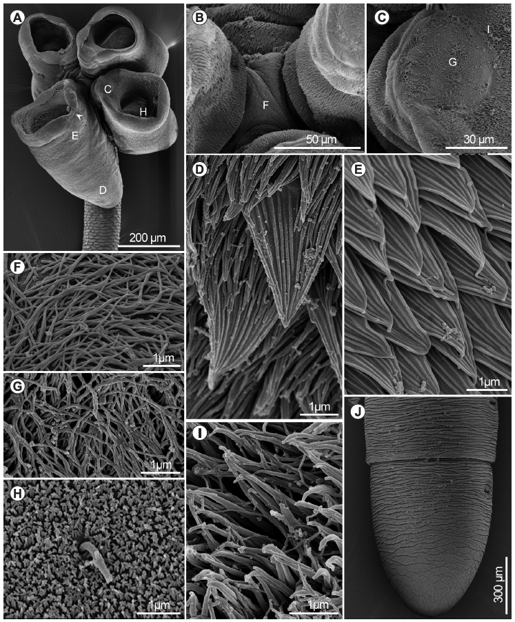 Fig. 6. Guidus magellanicus sp. n. from Bathyraja magellanica (Philippi), scanning electron micrographs. A scolex, small letters indicate locations of detail shown in CE and H, arrow indicates marginal indentation; B partial apical view of scolex showing bothridial stalks, small letter indicates location of detail shown in F; C apical sucker, small letter indicates location of detail shown in G; D proximal bothridial surface near proximal extreme, enlarged wide aristate spinitriches and capilliform filitriches; E proximal bothridial surface near bothridial rim, wide aristate spinitriches densely packed; F apex of scolex proper, capilliform filitriches; G surface of apical sucker, capilliform filitriches; H distal bothridial surface near base of internal cavity, acicular filitriches and cilium; I bothridial rim, narrow gladiate spinitriches and capilliform filitriches; J terminal portion of strobila showing scutes

