Line Drawing 1
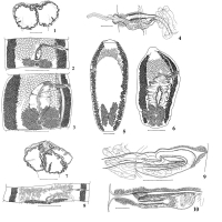
From Vasileva etal, 2002, Cit# 6149; Figures 1-3. Phyllobothrium squali Yamaguti, 1952, specimens from Squalus acanthias from Bulgaria. 1. Scolex. 2. Young mature proglottis.
3.Well-developed mature ... MoreFrom Vasileva etal, 2002, Cit# 6149; Figures 1-3. Phyllobothrium squali Yamaguti, 1952, specimens from Squalus acanthias from Bulgaria. 1. Scolex. 2. Young mature proglottis.
3.Well-developed mature proglottis. In Figures 2 and 3, the dorsal half of poral vitelline band is not shown in order to present genital ducts and
osmoregulatory canals. Scale-bars: 1 mm. Figures 4-6. Phyllobothrium squali Yamaguti, 1952, specimens from Squalus acanthias from Bulgaria. 4. Terminal genital ducts. 5. Gravid
proglottis, dorsal view. 6. Gravid proglottides, ventral view. In Figures 5 and 6, parts of poral vitelline band are not shown. Scale-bars: 4,
200 μm; 5,6, 1 mm. Figures 7-10. Phyllobothrium squali Yamaguti, 1952, holotype. 7. Scolex. 8. Pre-mature proglottis. 9. Terminal genital ducts. 10. Transverse
section through terminal genital ducts in mature proglottis. Scale-bars: 7, 1 mm; 8, 300 μm; 9, 150 μm; 10, 100 μm. |
Line Drawing 2

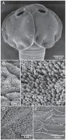
From Vasileva etal, 2002, Cit# 6149; Figure 11. Phyllobothrium squali Yamaguti, 1952, holotype.
Pre-gravid proglottis. Dorsal half of the poral vitelline band not
shown. Scale-bar: 500 μm. |
Photo Micrograph
|
Scanning Electron Micrograph
From Caira etal 2021 (Cit #7240); Figure 5. Scanning electron micrographs of Yamaguticestus. A, scolex of Yamaguticestus squali. B, surface of bothridium of Y. squali anterior to apical sucker. C, dis... MoreFrom Caira etal 2021 (Cit #7240); Figure 5. Scanning electron micrographs of Yamaguticestus. A, scolex of Yamaguticestus squali. B, surface of bothridium of Y. squali anterior to apical sucker. C, distal surface of bothridium of Y. squali. D, proximal surface of bothridium of Y. squali. E, scutes on surface of neck of Y. squali. |
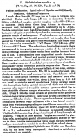
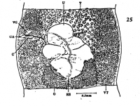

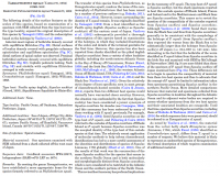
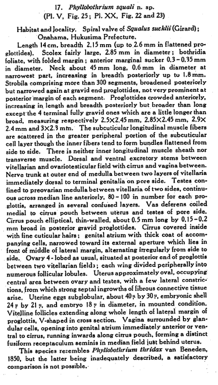
 PLATE V. FIGURE 25. Phyllobothrium squali n. sp. 25. Gravid proglottis of Phyllobothrium squali, dorsal view.
 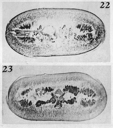 PLATE XX. FIGURES 22 & 23. Phyllobothrium squali n. sp. 22. Transverse section of not yet fully mature proglottis of Phyllobothrium squali through cirrus pouch and vagina, 25x. 23. Same through ovary, 25x.
  From Vasileva etal, 2002, Cit# 6149; Figures 1-3. Phyllobothrium squali Yamaguti, 1952, specimens from Squalus acanthias from Bulgaria. 1. Scolex. 2. Young mature proglottis.
3.Well-developed mature proglottis. In Figures 2 and 3, the dorsal half of poral vitelline band is not shown in order to present genital ducts and
osmoregulatory canals. Scale-bars: 1 mm. Figures 4-6. Phyllobothrium squali Yamaguti, 1952, specimens from Squalus acanthias from Bulgaria. 4. Terminal genital ducts. 5. Gravid
proglottis, dorsal view. 6. Gravid proglottides, ventral view. In Figures 5 and 6, parts of poral vitelline band are not shown. Scale-bars: 4,
200 μm; 5,6, 1 mm. Figures 7-10. Phyllobothrium squali Yamaguti, 1952, holotype. 7. Scolex. 8. Pre-mature proglottis. 9. Terminal genital ducts. 10. Transverse
section through terminal genital ducts in mature proglottis. Scale-bars: 7, 1 mm; 8, 300 μm; 9, 150 μm; 10, 100 μm.
From Vasileva etal, 2002, Cit# 6149; Figures 1-3. Phyllobothrium squali Yamaguti, 1952, specimens from Squalus acanthias from Bulgaria. 1. Scolex. 2. Young mature proglottis.
3.Well-developed mature proglottis. In Figures 2 and 3, the dorsal half of poral vitelline band is not shown in order to present genital ducts and
osmoregulatory canals. Scale-bars: 1 mm. Figures 4-6. Phyllobothrium squali Yamaguti, 1952, specimens from Squalus acanthias from Bulgaria. 4. Terminal genital ducts. 5. Gravid
proglottis, dorsal view. 6. Gravid proglottides, ventral view. In Figures 5 and 6, parts of poral vitelline band are not shown. Scale-bars: 4,
200 μm; 5,6, 1 mm. Figures 7-10. Phyllobothrium squali Yamaguti, 1952, holotype. 7. Scolex. 8. Pre-mature proglottis. 9. Terminal genital ducts. 10. Transverse
section through terminal genital ducts in mature proglottis. Scale-bars: 7, 1 mm; 8, 300 μm; 9, 150 μm; 10, 100 μm.  From Vasileva etal, 2002, Cit# 6149; Figure 11. Phyllobothrium squali Yamaguti, 1952, holotype.
Pre-gravid proglottis. Dorsal half of the poral vitelline band not
shown. Scale-bar: 500 μm.
From Vasileva etal, 2002, Cit# 6149; Figure 11. Phyllobothrium squali Yamaguti, 1952, holotype.
Pre-gravid proglottis. Dorsal half of the poral vitelline band not
shown. Scale-bar: 500 μm. 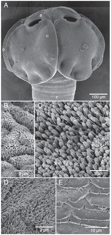 From Caira etal 2021 (Cit #7240); Figure 5. Scanning electron micrographs of Yamaguticestus. A, scolex of Yamaguticestus squali. B, surface of bothridium of Y. squali anterior to apical sucker. C, distal surface of bothridium of Y. squali. D, proximal surface of bothridium of Y. squali. E, scutes on surface of neck of Y. squali.
  From Caira etal 2021, Cit #7240

