Line Drawing 1

FIGURE 1 Echeneibothrium oligotesticularis sp. nov. a. Holotype - entire worm. b. A proglottid. c. Transverse section showing structure of body wall. Abbreviations: c, cirrus inside pouch; c.m., circ... MoreFIGURE 1 Echeneibothrium oligotesticularis sp. nov. a. Holotype - entire worm. b. A proglottid. c. Transverse section showing structure of body wall. Abbreviations: c, cirrus inside pouch; c.m., circular muscles; cu., cuticle; ex., excretory vessels in the sub-cuticular layer; l.m., longitudinal muscle bundles; ov., ovary; r.s., receptacleum seminis; s.gl., shell gland; t., testis; ut.,uterus; v., vagina; v.d., vas deferens; v.gl., vitelline glands; v.w., wall of cirrus pouch. |
Line Drawing 2

FIGURE 2. Echeneibothrium oligotesticularis sp. nov. a. Transverse section passing through the commissure of the ovary. b. Transverse section passing through shell gland. Abbreviations: c.c., centra... MoreFIGURE 2. Echeneibothrium oligotesticularis sp. nov. a. Transverse section passing through the commissure of the ovary. b. Transverse section passing through shell gland. Abbreviations: c.c., central commissure of ovary; d. ov., dorsal lobe of ovary; ex. d., main excretory duct; l.m., longitudinal muscle bundles; s.gl., shell gland; ut.,uterus; v., vagina; v.d., vas deferens; v. d1., vitelline duct; v.gl., vitelline glands; v. ov., ventral lobe of ovary. |
Photo Micrograph
|
Scanning Electron Micrograph
|

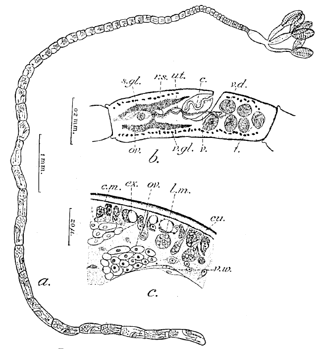 FIGURE 1 Echeneibothrium oligotesticularis sp. nov. a. Holotype - entire worm. b. A proglottid. c. Transverse section showing structure of body wall. Abbreviations: c, cirrus inside pouch; c.m., circular muscles; cu., cuticle; ex., excretory vessels in the sub-cuticular layer; l.m., longitudinal muscle bundles; ov., ovary; r.s., receptacleum seminis; s.gl., shell gland; t., testis; ut.,uterus; v., vagina; v.d., vas deferens; v.gl., vitelline glands; v.w., wall of cirrus pouch.
  FIGURE 2. Echeneibothrium oligotesticularis sp. nov. a. Transverse section passing through the commissure of the ovary. b. Transverse section passing through shell gland. Abbreviations: c.c., central commissure of ovary; d. ov., dorsal lobe of ovary; ex. d., main excretory duct; l.m., longitudinal muscle bundles; s.gl., shell gland; ut.,uterus; v., vagina; v.d., vas deferens; v. d1., vitelline duct; v.gl., vitelline glands; v. ov., ventral lobe of ovary.