Line Drawing 1
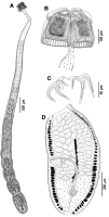
Fig. 1 Phoreiobothrium sorrahcola n. sp. a Whole worm, b scolex, c
hooks, d mature proglottid |
Line Drawing 2
|
Photo Micrograph
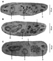
Fig. 3 Cross-sections through
proglottids of Phoreiobothrium
sorrahcola n. sp. a Cross-
section through testes anterior
to cirrus sac, b cross-section
through cirrus sac, c cross-
section throug... MoreFig. 3 Cross-sections through
proglottids of Phoreiobothrium
sorrahcola n. sp. a Cross-
section through testes anterior
to cirrus sac, b cross-section
through cirrus sac, c cross-
section through bilobed ovary.
cs cirrus sac, o ovary, t testis, u
uterus, v vagina, vi vitelline |
Scanning Electron Micrograph

Fig. 2 Surface ultrastructure of Phoreiobothrium sorrahcola n. sp.
a Scolex, b double septum between anterior and posterior loculi, c posterior part of anterior loculus and posterior loculus with its... MoreFig. 2 Surface ultrastructure of Phoreiobothrium sorrahcola n. sp.
a Scolex, b double septum between anterior and posterior loculi, c posterior part of anterior loculus and posterior loculus with its subloculi,
d papilliform to acicular filitriches on anterior muscular pad, e papilliform to acicular filitriches on proximal bothridial surface, f papilliform to acicular filitriches on anterior loculus, g papilliform to acicular filitriches on boundary between anterior and posterior loculus, h papilliform to acicular filitriches on posterior loculus, i cephalic peduncle with gladiate spinitriches interspersed with papilliform to acicular filitriches, j papilliform to acicular filitriches on cephalic peduncle, k proglottid with gladiate spinitriches interspersed with
papilliform to acicular filitriches, l papilliform to acicular filitriches
on proglottid. Note that small letters in a indicate locations of details shown in dj |
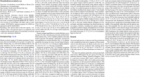
 Fig. 1 Phoreiobothrium sorrahcola n. sp. a Whole worm, b scolex, c
hooks, d mature proglottid
Fig. 1 Phoreiobothrium sorrahcola n. sp. a Whole worm, b scolex, c
hooks, d mature proglottid  Fig. 3 Cross-sections through
proglottids of Phoreiobothrium
sorrahcola n. sp. a Cross-
section through testes anterior
to cirrus sac, b cross-section
through cirrus sac, c cross-
section through bilobed ovary.
cs cirrus sac, o ovary, t testis, u
uterus, v vagina, vi vitelline
Fig. 3 Cross-sections through
proglottids of Phoreiobothrium
sorrahcola n. sp. a Cross-
section through testes anterior
to cirrus sac, b cross-section
through cirrus sac, c cross-
section through bilobed ovary.
cs cirrus sac, o ovary, t testis, u
uterus, v vagina, vi vitelline 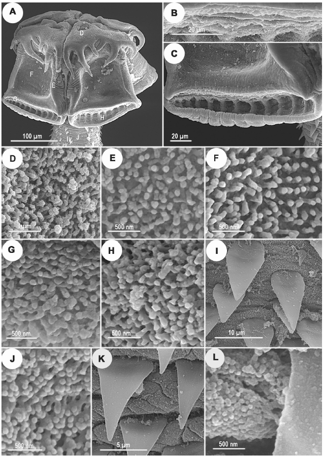 Fig. 2 Surface ultrastructure of Phoreiobothrium sorrahcola n. sp.
a Scolex, b double septum between anterior and posterior loculi, c posterior part of anterior loculus and posterior loculus with its subloculi,
d papilliform to acicular filitriches on anterior muscular pad, e papilliform to acicular filitriches on proximal bothridial surface, f papilliform to acicular filitriches on anterior loculus, g papilliform to acicular filitriches on boundary between anterior and posterior loculus, h papilliform to acicular filitriches on posterior loculus, i cephalic peduncle with gladiate spinitriches interspersed with papilliform to acicular filitriches, j papilliform to acicular filitriches on cephalic peduncle, k proglottid with gladiate spinitriches interspersed with
papilliform to acicular filitriches, l papilliform to acicular filitriches
on proglottid. Note that small letters in a indicate locations of details shown in dj
Fig. 2 Surface ultrastructure of Phoreiobothrium sorrahcola n. sp.
a Scolex, b double septum between anterior and posterior loculi, c posterior part of anterior loculus and posterior loculus with its subloculi,
d papilliform to acicular filitriches on anterior muscular pad, e papilliform to acicular filitriches on proximal bothridial surface, f papilliform to acicular filitriches on anterior loculus, g papilliform to acicular filitriches on boundary between anterior and posterior loculus, h papilliform to acicular filitriches on posterior loculus, i cephalic peduncle with gladiate spinitriches interspersed with papilliform to acicular filitriches, j papilliform to acicular filitriches on cephalic peduncle, k proglottid with gladiate spinitriches interspersed with
papilliform to acicular filitriches, l papilliform to acicular filitriches
on proglottid. Note that small letters in a indicate locations of details shown in dj 

