Cestode Scientific Name
| Species ID | 14162 |
|---|---|
| Order | Phyllobothriidea |
| Family | Phyllobothriidae |
| Subfamily | |
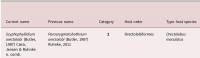
| Genus | Scyphophyllidium |
| Species | orectolobi |
| Authority | (Butler, 1987) Caira, Jensen & Ruhnke, 2020 |
| Taxonomic Status | Valid |
| Valid Name | |
| Synonyms | Phyllobothrium orectolobi Butler, 1987; Paraorygmatobothrium orectolobi (Butler, 1987) Ruhnke, 2011 |
| Genus Record | No |
| Type Species | No |
| Verified | Yes |
| Verified By | T. R. Ruhnke |
| Citation(s) |
Butler, S. A. 1987. Taxonomy of some tetraphyllidean cestodes from elasmobranch fishes. Australian Journal of Zoology 35: 343-371. (85) Download PDFCaira, J. N., K. Jensen, C. Hayes and T. R. Ruhnke. 2020. Insights from new cestodes of the crocodile shark, Pseudocarcharias kamoharai (Lamniformes: Pseudocarchariidae), prompt expansion of Scyphophyllidium and syonymization of seven phyllobothriidean generaat last!. Journal of Helminthology (https://doi.org/10.1017/S0022149X20000036): 124. (7164) Download PDF |
| Redescription |
Ruhnke, T. R. 2011. Tapeworms of Elasmobranchs (Part III). A monograph on the Phyllobothriidae (Platyhelminthes: Cestoda). Bulletin of the University of Nebraska State Museum 25: 205 pp. (5498) Download PDF |
| Scientific Name Notes | Ruhnke, 2011 (Cit #5498) includes a modified description |
Record Data
| Date (MM/DD/YYYY) | Action | User Name |
|---|---|---|
| 07/13/2020 | Created | B. Barbeau |
| 07/13/2020 | Modified | B. Barbeau |
| 07/15/2020 | Modified | B. Barbeau |
Type Host
| Host Class | |||||||
|---|---|---|---|---|---|---|---|
| Host Order | Orectolobiformes | ||||||
| Host Family | Orectolobidae | ||||||
|
Type Host (Literal) |
|
||||||
|
Type Host (Valid) |
|
||||||
| Additional Host(s) | |||||||
| Site in Host | |||||||
| Host Notes |
Type Locality
| Country | Australia |
|---|---|
| Body of Water | Moreton Bay |
| Island(s) | |
| City/Region | Queensland |
| Coordinates | |
| DD Latitude | |
| DD Longitude | |
| Additional Localities | |
| Locality Notes |
Specimens
| Type Material | QM No. GL4617 (holotype) QM No. GL4618-4620 (paratypes) |
|---|---|
| Total Number of Type Specimens | 25 |
| Voucher Material | |
| Specimen Notes |
Data are given as in original description unless otherwise indicated.
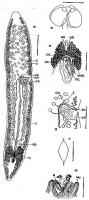

 FIGURES 15-20. Phyllobothrium orectolobi n. sp. 15. Scolex. 16. Gravid segment, ventral view. 17. Egg capsule. 18. Male reproductive system, ventral view, showing arrangement of collecting ducts. 19. Female reproductive system, ventral view. 20. Attached scolex. Abbreviations: b, bothridium; cc, copulatory canal; cd, collecting duct; cp, cirrus pouch; gp, genital pore; mu, mucosa; mvd, median vitelline duct; o, ovary; oc, oocapt; oo, ootype; ov, oviduct; sr, seminal receptacle; sv, seminal vessicle; t, testis; u, uterus; ud, uterine duct; v, vitellaria. Scale bars: 15, 16, 20, 0.44 mm; 17, 50 µm; 18 & 19, 0.2 mm.
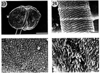
  FIGURES 23-26. Phyllobothrium orectolobi n. sp. 23. Scolex. 24. Anterior strobila; note scaly appearance. 25. Attaching surface of bothridium; note microvilli. 26. Posterior surface of bothridium; note blade-like projections. Scale bars: 23, 200 µm; 24, 20 µm; 25, 10 µm; 26, 4 µm.
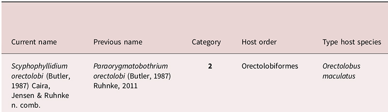
Best viewed in Firefox


