Cestode Scientific Name
| Species ID | 14065 |
|---|---|
| Order | Trypanorhyncha |
| Family | |
| Subfamily | |
| Genus | Nybelinia |
| Species | exostigmi |
| Authority | Quraishy, Dkhil, Abdel-Gaber, Al-Shaebi, Jaffal and Morsy, 2019 |
| Taxonomic Status | Unavailable name |
| Valid Name | |
| Synonyms | |
| Genus Record | No |
| Type Species | No |
| Verified | No |
| Verified By | |
| Citation(s) |
Quraishy, S. A., M. A. M Dkhil, R. Abdel-Gaber, E. Al-Shaebi, A. A. Jaffal, K. Morsy. 2019. Morphological and molecular insights of a new species of trypanorhynchid cestode parasite, Nybelinia exostigmi, in the Narrowstripe cardinal fish Apogon exostigma. Brazilian Journal of Veterinary Parasitology 28(2): 266282. (7133) Download PDF |
| Redescription | |
| Scientific Name Notes | No type specimens designated or deposited; ~840 bp of 18S rRNA deposited in GenBank (MK084750.1); only molecular comparison made to justify novelty |
Record Data
| Date (MM/DD/YYYY) | Action | User Name |
|---|---|---|
| 08/06/2019 | Created | K. Herzog |
| 08/06/2019 | Modified | K. Herzog |
Type Host
| Host Class | Actinopterygii | ||||||
|---|---|---|---|---|---|---|---|
| Host Order | Perciformes | ||||||
| Host Family | Apogonidae | ||||||
|
Type Host (Literal) |
|
||||||
|
Type Host (Valid) |
|
||||||
| Additional Host(s) | |||||||
| Site in Host | alimentary canal | ||||||
| Host Notes | found in intestine and stomach; prevalence: 25 out of 40 (62.5%) specimens infected |
Type Locality
| Country | Egypt |
|---|---|
| Body of Water | Red Sea, Gulf of Suez |
| Island(s) | |
| City/Region | Hurghada |
| Coordinates | |
| DD Latitude | |
| DD Longitude | |
| Additional Localities | |
| Locality Notes |
Specimens
| Type Material | none |
|---|---|
| Total Number of Type Specimens | |
| Voucher Material | |
| Specimen Notes |
Data are given as in original description unless otherwise indicated.


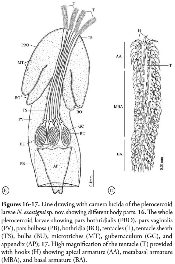 Figures 16-17. Line drawing with camera lucida of the plerocercoid larvae N. exostigmi sp. nov. showing different body parts. 16. The whole plerocercoid larvae showing pars bothridialis (PBO), pars vaginalis (PV), pars bulbosa (PB), bothridia (BO), tentacles (T), tentacle sheath (TS), bulbs (BU), microtriches (MT), gubernaculum (GC), and appendix (AP); 17. High magnification of the tentacle (T) provided with hooks (H) showing apical armature (AA), metabasal armature (MBA), and basal armature (BA).
 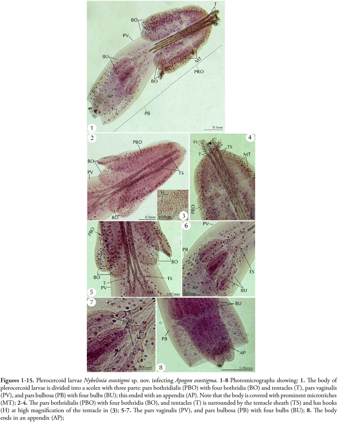 Figures 1-15. Plerocercoid larvae Nybelinia exostigmi sp. nov. infecting Apogon exostigma. 1-8 Photomicrographs showing: 1. The body of plerocercoid larvae is divided into a scolex with three parts: pars bothridialis (PBO) with four bothridia (BO) and tentacles (T), pars vaginalis (PV), and pars bulbosa (PB) with four bulbs (BU); this ended with an appendix (AP). Note that the body is covered with prominent microtriches (MT); 2-4. The pars bothridialis (PBO) with four bothridia (BO), and tentacles (T) is surrounded by the tentacle sheath (TS) and has hooks (H) at high magnification of the tentacle in (3); 5-7. The pars vaginalis (PV), and pars bulbosa (PB) with four bulbs (BU); 8. The body ends in an appendix (AP);
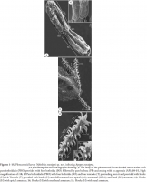
 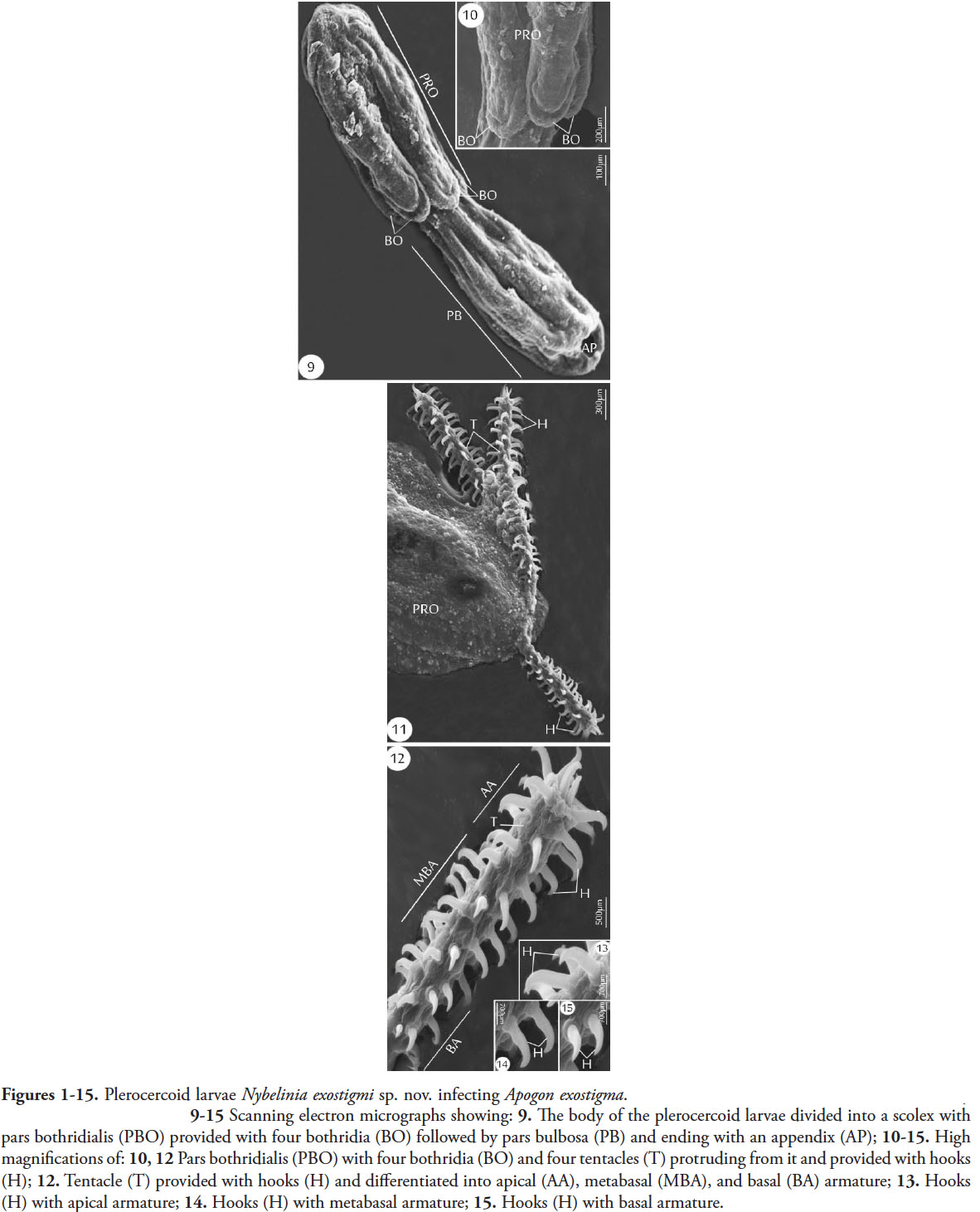 Figures 1-15. Plerocercoid larvae Nybelinia exostigmi sp. nov. infecting Apogon exostigma. 9-15 Scanning electron micrographs showing: 9. The body of the plerocercoid larvae divided into a scolex with
pars bothridialis (PBO) provided with four bothridia (BO) followed by pars bulbosa (PB) and ending with an appendix (AP); 10-15. High magnifications of: 10, 12 Pars bothridialis (PBO) with four bothridia (BO) and four tentacles (T) protruding from it and provided with hooks (H); 12. Tentacle (T) provided with hooks (H) and differentiated into apical (AA), metabasal (MBA), and basal (BA) armature; 13. Hooks (H) with apical armature; 14. Hooks (H) with metabasal armature; 15. Hooks (H) with basal armature.
Figures 1-15. Plerocercoid larvae Nybelinia exostigmi sp. nov. infecting Apogon exostigma. 9-15 Scanning electron micrographs showing: 9. The body of the plerocercoid larvae divided into a scolex with
pars bothridialis (PBO) provided with four bothridia (BO) followed by pars bulbosa (PB) and ending with an appendix (AP); 10-15. High magnifications of: 10, 12 Pars bothridialis (PBO) with four bothridia (BO) and four tentacles (T) protruding from it and provided with hooks (H); 12. Tentacle (T) provided with hooks (H) and differentiated into apical (AA), metabasal (MBA), and basal (BA) armature; 13. Hooks (H) with apical armature; 14. Hooks (H) with metabasal armature; 15. Hooks (H) with basal armature. Best viewed in Firefox


